Review
. 2021 Jul 28;22(15):8099.
doi: 10.3390/ijms22158099.

Unique Aspects of Human Placentation

Affiliations
Review

Unique Aspects of Human Placentation

Anthony M Carter. Int J Mol Sci. .

Abstract

Human placentation differs from that of other mammals. A suite of characteristics is shared with haplorrhine primates, including early development of the embryonic membranes and placental hormones such as chorionic gonadotrophin and placental lactogen. A comparable architecture of the intervillous space is found only in Old World monkeys and apes. The routes of trophoblast invasion and the precise role of extravillous trophoblast in uterine artery transformation is similar in chimpanzee and gorilla. Extended parental care is shared with the great apes, and though human babies are rather helpless at birth, they are well developed (precocial) in other respects. Primates and rodents last shared a common ancestor in the Cretaceous period, and their placentation has evolved independently for some 80 million years. This is reflected in many aspects of their placentation. Some apparent resemblances such as interstitial implantation and placental lactogens are the result of convergent evolution. For rodent models such as the mouse, the differences are compounded by short gestations leading to the delivery of poorly developed (altricial) young.

Keywords: decidual reaction; fetal membranes; placental hormones; primates; uterine NK cell; uterine spiral artery.


Conflict of interest statement

The author declares no conflict of interest.

Figures
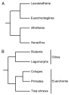
Figure 1
Figure 1
The mammalian tree. (A) The four major clades of eutherians [8]. (B) The orders of Euarchontoglires [9]. Note the separation of Glires (including rodents) from Euarchonta (including primates). There are alternative interpretations of the root of the tree and the position of tree shrews. Reprinted with permission from [2] © 2021 Society for Reproduction and Fertility.
Figure 2
Figure 2
Classification of primates [12]. Strepsirrhines and haplorrhines are suborders, tarsiers are regarded as an infraorder, whilst the other clades shown are superfamilies. OW, Old World; NW, New World. Reprinted with permission from [2] © 2021 Society for Reproduction and Fertility.
Figure 3
Figure 3
Early differentiation of mesoderm in the human embryo. (a) Trophoblastic plate stage (Carnegie Stage 5a) showing the inner cell mass (icm). The cavity of the blastocyst is collapsed. The pad of trophoblast has different sized nuclei and both cellular and syncytial trophoblast (syn tr) (Carnegie Embryo #8020). Scale bar, 70 µm. (b) Early lacunar stage (Carnegie Stage 5b) Some maternal blood has leaked into the primary yolk sac (pys). Note the irregular shape of the lacunae on the right, which appear to be formed from expanding clefts (Carnegie Embryo #8004). Scale bar, 90 µm. (c) Lacunar stage (Carnegie Stage 5c). Note the anastomotic lacunae within the syncytiotrophoblast. In the area between the trophoblast and the already partially constricted primary yolk sac (pys), there are mesenchymal cells (extraembryonic mesoderm) (Carnegie Embryo #7699). Scale bar, 176 µm. (d) Predecessors of the primary villi (Carnegie Stage 6). The cytotrophoblast (cyt troph) is accumulating in the partitions between lacunae, initiating the formation of primary villi (Carnegie Embryo #9260). Scale bar = 70 µm. Reprinted with permission from Carter, Enders and Pijnenborg [16] © The Authors. Published by the Royal Society. All rights reserved.
Figure 4
Figure 4
The interhaemal barrier of the human placenta is classified as haemomonochorial. The intervillous space is separated from blood in the fetal capillary by syncytiotrophoblast and fetal capillary endothelium with their basal membranes. A very thin layer of connective tissue cytoplasm is interposed between the two basal membranes. Courtesy of Dr. Allen C. Enders. Reproduced with permission from [35] Copyright © American Physiological Society.

References

    1. Brosens I., Pijnenborg R., Vercruysse L., Romero R. The “Great Obstetrical Syndromes” are associated with disorders of deep placentation. Am. J. Obstet. Gynecol. 2011;204:193–201. doi: 10.1016/j.ajog.2010.08.009. - DOI - PMC - PubMed
    1. Carter A.M. Animal models of human pregnancy and placentation: Alternatives to the mouse. Reproduction. 2020;160:R129–R143. doi: 10.1530/REP-20-0354. - DOI - PubMed
    1. Carter A.M. Animal models of human placentation—A review. Placenta. 2007;28:S41–S47. doi: 10.1016/j.placenta.2006.11.002. - DOI - PubMed
    1. Carter A.M. Evolution of Placental Function in Mammals: The Molecular Basis of Gas and Nutrient Transfer, Hormone Secretion, and Immune Responses. Physiol. Rev. 2012;92:1543–1576. doi: 10.1152/physrev.00040.2011. - DOI - PubMed
    1. Huhn O., Zhao X., Esposito L., Moffett A., Colucci F., Sharkey A.M. How Do Uterine Natural Killer and Innate Lymphoid Cells Contribute to Successful Pregnancy? Front. Immunol. 2021;12:607669. doi: 10.3389/fimmu.2021.607669. - DOI - PMC - PubMed

Substances

LinkOut - more resources