. 2021 Jul 29;22(15):8143.
doi: 10.3390/ijms22158143.

Computational Analysis of the Crystal and Cryo-EM Structures of P-Loop Channels with Drugs

Affiliations

Computational Analysis of the Crystal and Cryo-EM Structures of P-Loop Channels with Drugs

Denis B Tikhonov et al. Int J Mol Sci. .

Abstract

The superfamily of P-loop channels includes various potassium channels, voltage-gated sodium and calcium channels, transient receptor potential channels, and ionotropic glutamate receptors. Despite huge structural and functional diversity of the channels, their pore-forming domain has a conserved folding. In the past two decades, scores of atomic-scale structures of P-loop channels with medically important drugs in the inner pore have been published. High structural diversity of these complexes complicates the comparative analysis of these structures. Here we 3D-aligned structures of drug-bound P-loop channels, compared their geometric characteristics, and analyzed the energetics of ligand-channel interactions. In the superimposed structures drugs occupy most of the sterically available space in the inner pore and subunit/repeat interfaces. Cationic groups of some drugs occupy vacant binding sites of permeant ions in the inner pore and selectivity-filter region. Various electroneutral drugs, lipids, and detergent molecules are seen in the interfaces between subunits/repeats. In many structures the drugs strongly interact with lipid and detergent molecules, but physiological relevance of such interactions is unclear. Some eukaryotic sodium and calcium channels have state-dependent or drug-induced π-bulges in the inner helices, which would be difficult to predict. The drug-induced π-bulges may represent a novel mechanism of gating modulation.

Keywords: 3D alignment; Monte Carlo minimization; ligand-receptor interactions; sequence alignment; π-bulges.


Conflict of interest statement

The authors declare no conflict of interest.

Figures

Figure 1
Figure 1
Sequence alignment of helices in the pore domain of P-loop channels. Universal labels of residues are shown above the sequences.
Figure 2
Figure 2
Ligands in the pore domain of P-loop channels. Carbon, oxygen and nitrogen atoms of ligands are gray, red and blue, respectively. (A), Cα tracing of the pore domain in 25 drug-bound P-loop channels. For clarity, only two subunits/repeats without drugs are shown. The structures are 3D-aligned as described in Methods. (B), Superimposition of ligand-channel complexes with ligands in the inner pore. Only two subunits of the KcsA channels are shown for clarity. Ligand molecules fill the entire central cavity. (C), Superimposition of channel complexes with ligands bound in subunit/repeat interfaces (between P1 helix and two inner helices). Most ligands are close to the pore. Exceptions are DHP drugs in CavAb, which are far from the pore axis. (D), Lipid and detergent molecules bind in the inner-pore central cavity, fenestrations, or in the activation gate region.
Figure 3
Figure 3
KcsA pore domain (green helices) with superimposed ligands from KcsA, GluR, NavAb, and CavAb channels. (A,B), Intracellular and intra-membrane views of the KcsA complex with tetradecylammonium (PDB ID: 2w0f). The ammonium nitrogen is at the focus of P-helices. Long decyl chains penetrate into subunit interfaces and reach the outer helices. Main channel contributors to the ligand-binding energy are shown by sticks. (C), 3D-aligned structures of TBA-bound KcsA (PDB ID: 2hvj) (green backbone and ligand carbons), MK801-bound NMDAR channel (PDB ID: 5un1) (orange backbone and ligand carbons), and IEM-1460 bound AMPAR channel (PDB ID: 6dm0) (cyan backbone and ligand carbons). The central cavity of the channels accommodates bulky moieties of TBA, MK801, and IEM-1460. Magenta spheres show potassium ions in Cd2+-bound KcsA (PDB ID: 3stl). The terminal ammonium group of IEM-1460 penetrates into the selectivity filter and binds near Site 3 of potassium ion; the second ammonium nitrogen binds close to the position of TBA ammonium nitrogen in KcsA. The ammonium group of MK801 binds in the central cavity, close to Site 5 of potassium ion KcsA. (D), Ammonium nitrogen atoms of cationic drugs (blue spheres) scatter over the central cavity.
Figure 4
Figure 4
π-Bugles in the inner helices of P-loop channels. (A), Intracellular and membrane vies at superimposition of KcsA (PDB ID: 1bl8), Kv1.2/Kv2.1 (PDB ID: 2r9r), AMPAR (PDB ID: 6dm0), NavAb (PDB ID: 3rvy), NavMs (PDB ID: 6yz0), and GsuK (PDB ID: 4gx5). CA-CB bonds in positions i9 and i20 are shown as sticks. Upstream from the π-bulge (position i9), orientations of CA-CB bonds (gray CA, pink CB) are similar, while downstream the bulge (i20 position), orientation of CA-CB bonds in GsuK (gray CA, red CB) is different from that in other channels. (B), Deviations of alpha carbons in representative structures of P-loop channels from sequentially matching positions in the KcsA structure (PDB ID: 1bl8). Another KcsA structure (PDB ID: 2boc) has small deviations. For the open-gate Kv1.2/Kv2.1 channel, the deviations are large at the C-terminal part, but the deviation plot remains rather smooth. For the GsuK structures (PDB IDs: 4gx0, 4gx5), with a bulge in the middle of the inner helix, large oscillations are seen at the plots. (C), Distances of alpha-carbons in the inner helices from the pore axis. The bulge in the GsuK structures causes a shift of maxima and minima in the distance plot.
Figure 5
Figure 5
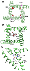
π-Bulges affect patterns of ligand-sensing residues in eukaryotic Nav channels. (A), Ni20_Cα-Cβ bonds in regular alpha-helices and π-bulged helices are shown with magenta and red CB atoms, respectively. B, A steroidal molecule in hNav1.4 (PDB ID: 6afg). F2i30 and F4i30 face the pore and interact with the steroid, while residues in sequentially matching positions, F3i30 and Y1i30 (Figure 1) face away from the pore. C, Flecainide in rNav1.5 (PDB ID: 6uz0). Residues F2i22 and L3i19 are oriented toward the pore-bound ligand, whereas F3i22 and L2i19 are oriented away from the pore.
Figure 6
Figure 6
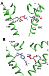
π-Bulges affect patterns of ligand-sensing residues in Cav1.1 channel. (A), Diverse orientation of asparagines Ni20. Orientation of Ni20_Cα-Cβ bonds in regular and π-bulged helices are shown with magenta and red CB atoms, respectively. (B), Different orientation of adjacent M3i18 and M3i19 in Cav1.1 Class II structure (PDB ID: 6jp5, magenta backbone, orange sidechains) and Class III structure (PDB ID: 6jpa; green backbone, cyan sidechains). M3i18 faces the DHP site in Class II structure, whereas M3i19 faces the DHP site in Class III structure. C, Superimposition of Cav3.1 structures with Z944 (6kzp) and without Z944 (6kzo). Residues in matching positions 2i19 and 2i22 differently interact with the ligand since the π-bulge is present only in 6kzp.
Figure 7
Figure 7
Examples of ligand interactions with the lipid molecules in the pore. (A), Amlodipine in Cav1.1 (PDB ID: 7jpx). The ammonium group approaches the outer pore and forms a salt bridge with a phospholipid molecule. (B), Z944 and lipid molecules fill up the cavity in Cav3.1 (PDB ID: 6kzp). The ligand amino group and phosphate group of the lipid (pink phosphorus) are salt-bridged.

Similar articles

Cited by

References

    1. Doyle D.A., Morais Cabral J., Pfuetzner R.A., Kuo A., Gulbis J.M., Cohen S.L., Chait B.T., MacKinnon R. The structure of the potassium channel: Molecular basis of K+ conduction and selectivity. Science. 1998;280:69–77. doi: 10.1126/science.280.5360.69. - DOI - PubMed
    1. Burley S.K., Berman H.M., Kleywegt G.J., Markley J.L., Nakamura H., Velankar S. Protein Data Bank (PDB): The single global macromolecular structure archive. Methods Mol. Biol. 2017;1607:627–641. doi: 10.1007/978-1-4939-7000-1_26. - DOI - PMC - PubMed
    1. Zhorov B.S., Tikhonov D.B. Potassium, sodium, calcium and glutamate-gated channels: Pore architecture and ligand action. J. Neurochem. 2004;88:782–799. doi: 10.1111/j.1471-4159.2004.02261.x. - DOI - PubMed
    1. Dong Y.Y., Pike A.C., Mackenzie A., McClenaghan C., Aryal P., Dong L., Quigley A., Grieben M., Goubin S., Mukhopadhyay S., et al. K2P channel gating mechanisms revealed by structures of TREK-2 and a complex with Prozac. Science. 2015;347:1256–1259. doi: 10.1126/science.1261512. - DOI - PMC - PubMed
    1. Tang L., Gamal El-Din T.M., Swanson T.M., Pryde D.C., Scheuer T., Zheng N., Catterall W.A. Structural basis for inhibition of a voltage-gated Ca(2+) channel by Ca(2+) antagonist drugs. Nature. 2016;537:117–121. doi: 10.1038/nature19102. - DOI - PMC - PubMed

MeSH terms