Review
. 2021 Jul 30;22(15):8180.
doi: 10.3390/ijms22158180.

Mitochondrial Modulations, Autophagy Pathways Shifts in Viral Infections: Consequences of COVID-19

Affiliations
Review

Mitochondrial Modulations, Autophagy Pathways Shifts in Viral Infections: Consequences of COVID-19

Shailendra Pratap Singh et al. Int J Mol Sci. .

Abstract

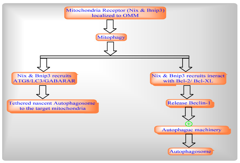
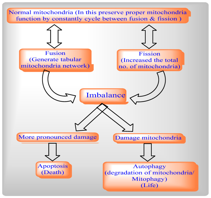
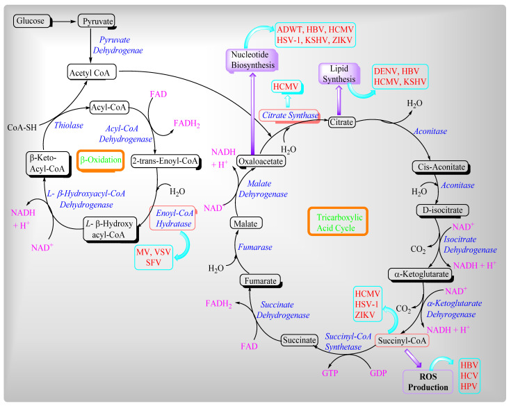
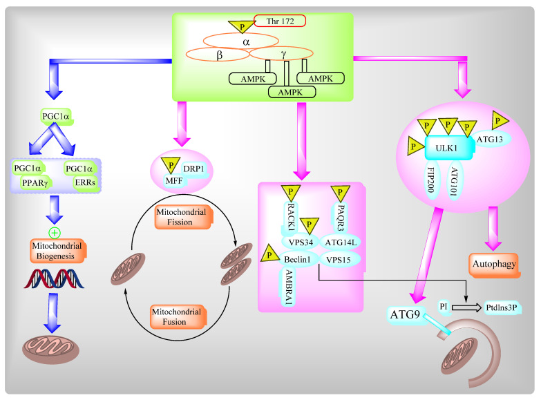
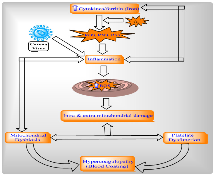
Mitochondria are vital intracellular organelles that play an important role in regulating various intracellular events such as metabolism, bioenergetics, cell death (apoptosis), and innate immune signaling. Mitochondrial fission, fusion, and membrane potential play a central role in maintaining mitochondrial dynamics and the overall shape of mitochondria. Viruses change the dynamics of the mitochondria by altering the mitochondrial processes/functions, such as autophagy, mitophagy, and enzymes involved in metabolism. In addition, viruses decrease the supply of energy to the mitochondria in the form of ATP, causing viruses to create cellular stress by generating ROS in mitochondria to instigate viral proliferation, a process which causes both intra- and extra-mitochondrial damage. SARS-COV2 propagates through altering or changing various pathways, such as autophagy, UPR stress, MPTP and NLRP3 inflammasome. Thus, these pathways act as potential targets for viruses to facilitate their proliferation. Autophagy plays an essential role in SARS-COV2-mediated COVID-19 and modulates autophagy by using various drugs that act on potential targets of the virus to inhibit and treat viral infection. Modulated autophagy inhibits coronavirus replication; thus, it becomes a promising target for anti-coronaviral therapy. This review gives immense knowledge about the infections, mitochondrial modulations, and therapeutic targets of viruses.

Keywords: COVID-19; SARS-COV2; autophagy; mitochondria; potential targets; viral infections.


Conflict of interest statement

The authors declare no conflict of interest.

Figures

Figure 1
Figure 1
—Steps of Autophagy: Autophagy (macro autophagy) means “self” (“auto”) and “eating” (“phagy”). Autophagy removes dysfunctional and unnecessary cellular components through various steps such as Omegasomes and the initiation of isolation membrane; the elongation of the isolation membrane and formation of autophagosomes; and autophagosome-lysosome fusion and degradation.
Figure 2
Figure 2
Types of Autophagy: Autophagy categorized into three different categories based on differences in their routes to the lysosome.
Figure 3
Figure 3
ER stress-induced unfolded protein receptor [UPR] pathways to cause Autophagy.
Figure 4
Figure 4
Autophagy pathway including the convergence of the endocytic pathways.
Figure 5
Figure 5
Damaged-Induced Mitophagy involves two major proteins: [1] ubiquitin kinase PINK1 (which flags the damaged mitochondria); and [2] parkin, E3 ligase (as a signal amplifier).
Figure 6
Figure 6
Development-Induced Mitophagy.
Figure 7
Figure 7
Relationships between mitochondrial fission and fusion, apoptosis and mitophagy.
Figure 8
Figure 8
Viruses’ influence on beta-oxidation and the TCA cycle.
Figure 9
Figure 9
AMPK regulates a variety of metabolic processes.
Figure 10
Figure 10
Mitochondria dysfunction in the pathogenesis of COVID-19.

References

    1. Bratic I., Trifunovic A. Mitochondrial energy metabolism and ageing. Biochim. Biophys. Acta (BBA) Bioenerg. 2010;1797:961–967. doi: 10.1016/j.bbabio.2010.01.004. - DOI - PubMed
    1. Westermann B. Mitochondrial fusion and fission in cell life and death. Nat. Rev. Mol. Cell Biol. 2010;11:872–884. doi: 10.1038/nrm3013. - DOI - PubMed
    1. James D.I., Parone P.A., Mattenberger Y., Martinou J.-C. hFis1, a Novel Component of the Mammalian Mitochondrial Fission Machinery. J. Biol. Chem. 2003;278:36373–36379. doi: 10.1074/jbc.M303758200. - DOI - PubMed
    1. Smirnova E., Shurland D.-L., Ryazantsev S.N., Van Der Bliek A.M. A Human Dynamin-related Protein Controls the Distribution of Mitochondria. J. Cell Biol. 1998;143:351–358. doi: 10.1083/jcb.143.2.351. - DOI - PMC - PubMed
    1. Zhu P.-P., Patterson A., Stadler J., Seeburg D.P., Sheng M., Blackstone C. Intra- and Intermolecular Domain Interactions of the C-terminal GTPase Effector Domain of the Multimeric Dynamin-like GTPase Drp1. J. Biol. Chem. 2004;279:35967–35974. doi: 10.1074/jbc.M404105200. - DOI - PubMed