. 2021 Jul 31;22(15):8241.
doi: 10.3390/ijms22158241.

Biocontrol of Biofilm Formation: Jamming of Sessile-Associated Rhizobial Communication by Rhodococcal Quorum-Quenching

Affiliations

Biocontrol of Biofilm Formation: Jamming of Sessile-Associated Rhizobial Communication by Rhodococcal Quorum-Quenching

Yvann Bourigault et al. Int J Mol Sci. .

Abstract

Biofilms are complex structures formed by a community of microbes adhering to a surface and/or to each other through the secretion of an adhesive and protective matrix. The establishment of these structures requires a coordination of action between microorganisms through powerful communication systems such as quorum-sensing. Therefore, auxiliary bacteria capable of interfering with these means of communication could be used to prevent biofilm formation and development. The phytopathogen Rhizobium rhizogenes, which causes hairy root disease and forms large biofilms in hydroponic crops, and the biocontrol agent Rhodococcus erythropolis R138 were used for this study. Changes in biofilm biovolume and structure, as well as interactions between rhizobia and rhodococci, were monitored by confocal laser scanning microscopy with appropriate fluorescent biosensors. We obtained direct visual evidence of an exchange of signals between rhizobia and the jamming of this communication by Rhodococcus within the biofilm. Signaling molecules were characterized as long chain (C14) N-acyl-homoserine lactones. The role of the Qsd quorum-quenching pathway in biofilm alteration was confirmed with an R. erythropolis mutant unable to produce the QsdA lactonase, and by expression of the qsdA gene in a heterologous host, Escherichia coli. Finally, Rhizobium biofilm formation was similarly inhibited by a purified extract of QsdA enzyme.

Keywords: N-acyl homoserine lactones; Rhizobium (Agrobacterium) rhizogenes; Rhodococcus erythropolis; biofilm; biological control; communication; hairy root; lifestyle switch; quorum-quenching; quorum-sensing.


Conflict of interest statement

The authors declare no conflict of interest.

Figures

Figure 1
Figure 1
HPLC–MS analyses of AHL signaling molecules in R. rhizogenes culture supernatants. The chromatogram of an AHL extract from strain 5520T (A1) and associated mass spectra of compounds from chromatogram peaks 1 (A2) and 2 (A3) were compared to the chromatogram of a mixture of two synthetic AHLs, 3-OH-C14:1-HSL and 3-OH-C14-HSL (B1), and the corresponding mass spectra of compounds from chromatogram peaks 3 (B2) and 4 (B3).
Figure 2
Figure 2
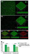
Inhibition of rhizobial biofilm formation by the biocontrol agent Rhodococcus erythropolis R138 (ratio 1:1). Biofilms were developed during incubation at 25 °C for 48 h in LB under static conditions. Confocal laser scanning microscopy (CLSM) analysis of the R. rhizogenes 5520T biofilm (A1) or dual-species biofilm formed by R. rhizogenes 5520T (green fluorescence) and R. erythropolis R138 (red fluorescence) (A2,A3) was achieved at an inoculation ratio of 1:1. R. rhizogenes and rhodococcal bacteria were tagged with green fluorescent protein (GFP) and mCherry via the pHC60-gfp and pEPR1-mCherry vectors, respectively. Top view, 3D shadow representation, and side view of the biofilm produced by R. rhizogenes alone (A1), and by a mixed culture of R. rhizogenes and R. erythropolis (A2) analyzed in the green channel. Location of rhodococcal cells was revealed by 3D shadow representation and side view of the biofilm produced by the mixed culture analyzed in the red plus green, green, or red channels (A3) (activation of the green or red channel is indicated by a light spot of the corresponding color). COMSTAT analyses of R. rhizogenes green fluorescence in single- and dual-species biofilms. R. rhizogenes biofilm biomass and thickness values were normalized (set to 100%) as a reference for a comparison with mixed biofilm conditions (B). The data shown are the means of three measurements from three independent experiments. Significant differences (Mann–Whitney test; p-value < 0.01) in biovolume and thickness are indicated by asterisks (★★★ p < 0.001).
Figure 3
Figure 3
Production of rhizobial AHL signaling molecules within the biofilm. CLSM analysis of the dual-species biofilm formed by R. rhizogenes 5520T and the P. atrosepticum 6267-EI AHL biosensor strain (1:1 ratio). Bacteria were labeled with the nucleic acid stain Syto 61 (bright red fluorescence), whereas the P. atrosepticum 6276-EI AHL-biosensor was also tagged by GFP, due to the gfp expression governed by the AHL inducible promoter. Top view, 3D shadow representation and side view of the biofilm produced by a mixed culture of R. rhizogenes and P. atrosepticum (A1,A2). We analyzed the structure of the biofilm (bright red fluorescence) due to the activation of the green plus red channels for (A1), and used the green channel only for (A2), for visualization of the presence of AHL (bright green fluorescence) in the biofilm through the expression of the gfp reporter gene under control of the AHL-inducible promoter (activation of the green or red channel is indicated by a light spot of the corresponding color).
Figure 4
Figure 4
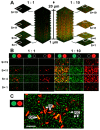
CLSM analysis of quorum-quenching activity in a dual-species biofilm of R. rhizogenes and R. erythropolis. R. rhizogenes (green fluorescence) was tagged with GFP via the pHC60-gfp. R. erythropolis was transformed with the pEPR1-qsdR-Pqsd::gfp-mCherry vector containing a transcriptional fusion between the promoter of the AHL lactonase-encoding gene qsdA and the gfp gene for the monitoring of its quorum-quenching activity. (A) A total of 19 confocal image stacks acquired with 1 µm slices from the bottom to the top of the mixed biofilms. For each mixed biofilm (with an R. rhizogenes/R. erythropolis ratio of 1:1 at left or 1:10 at right), four image stacks taken at 1 (S = 1), 4 (S = 4), 11 (S = 11), and 19 (S = 19) µm from the bottom of the biofilm were selected for the assessment of rhodococcal quorum-quenching activity. (B) Details delineated by the white circled area of four sample image stacks, previously selected among images in (A), to highlight the rhodococcal quorum-quenching activity at the different observation channels. (C) Close-up of the selected image stack S = 4 with an R. rhizogenes/R. erythropolis ratio of 1:1. Legend: RȻ, rhizobial cells (green fluorescence), QȻ, quenching cells sensing and degrading N-acyl-homoserine lactone (AHL) signals (green plus red, i.e., yellow, fluorescence); IȻ, inactive cell not sensing or degrading AHL signals (red fluorescence) (activation of the green or red channel is indicated by a light spot of the corresponding color). Scale bar represents 5 µm.
Figure 5
Figure 5
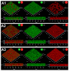
Effects of the R. erythropolis ΔqsdA deletion mutant and of QsdA AHL-lactonase-producing E. coli strains on R. rhizogenes biofilm formation. CLSM analysis of the dual-species biofilm formed by R. rhizogenes 5520T (green fluorescence) and R. erythropolis R138 (or E. coli) (red fluorescence) (1:1 ratio). R. rhizogenes and rhodococcal (or E. coli) bacteria were tagged with GFP and mCherry via the pHC60-gfp and pEPR1(or pUC19)-mCherry vectors, respectively. 3D representation and side view of the dual-species biofilm formed by R. rhizogenes and R. erythropolis ΔqsdA (A1), R. rhizogenes and E. coli pUC19-mCherry (A2), and R. rhizogenes and E. coli pUC19-qsdA-mCherry (A3) (activatation of the green or red channel is indicated by a light spot of the corresponding color). COMSTAT analysis of the biovolume and thickness of the biofilms developed. R. rhizogenes biofilm, biovolume, and thickness values were set to 100%, for use as a reference in comparison with mixed biofilm conditions (B). The data shown are the means of three measurements from three independent experiments. Significant differences in biovolume and thickness values (Mann–Whitney test; p-value < 0.01) are indicated by asterisks (★ p < 0.05); ns, not significant.
Figure 5
Figure 5
Effects of the R. erythropolis ΔqsdA deletion mutant and of QsdA AHL-lactonase-producing E. coli strains on R. rhizogenes biofilm formation. CLSM analysis of the dual-species biofilm formed by R. rhizogenes 5520T (green fluorescence) and R. erythropolis R138 (or E. coli) (red fluorescence) (1:1 ratio). R. rhizogenes and rhodococcal (or E. coli) bacteria were tagged with GFP and mCherry via the pHC60-gfp and pEPR1(or pUC19)-mCherry vectors, respectively. 3D representation and side view of the dual-species biofilm formed by R. rhizogenes and R. erythropolis ΔqsdA (A1), R. rhizogenes and E. coli pUC19-mCherry (A2), and R. rhizogenes and E. coli pUC19-qsdA-mCherry (A3) (activatation of the green or red channel is indicated by a light spot of the corresponding color). COMSTAT analysis of the biovolume and thickness of the biofilms developed. R. rhizogenes biofilm, biovolume, and thickness values were set to 100%, for use as a reference in comparison with mixed biofilm conditions (B). The data shown are the means of three measurements from three independent experiments. Significant differences in biovolume and thickness values (Mann–Whitney test; p-value < 0.01) are indicated by asterisks (★ p < 0.05); ns, not significant.
Figure 6
Figure 6
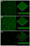
Effect of the purified AHL-lactonase QsdA on R. rhizogenes biofilm formation. Control conditions for biofilm formatin were established by substituting QsdA by bovine serum albumin (BSA). R. rhizogenes was transformed with the pHC60-gfp plasmid to tag bacteria by the constitutive expression of gfp (green fluorescent cells). Top view, 3D representation and side view of the R. rhizogenes biofilm in the presence of 100 µL of elution buffer alone (A1) or containing the BSA (A2) or the QsdA lactonase (A3). COMSTAT analysis of the biovolume and thickness of the biofilms developed (B). The data shown are the means of three measurements from three independent experiments. Significant differences in biovolume and thickness values (Mann–Whitney test; p-value < 0.01) are indicated by asterisks (★★ p < 0.01); ns, not significant.
Figure 6
Figure 6
Effect of the purified AHL-lactonase QsdA on R. rhizogenes biofilm formation. Control conditions for biofilm formatin were established by substituting QsdA by bovine serum albumin (BSA). R. rhizogenes was transformed with the pHC60-gfp plasmid to tag bacteria by the constitutive expression of gfp (green fluorescent cells). Top view, 3D representation and side view of the R. rhizogenes biofilm in the presence of 100 µL of elution buffer alone (A1) or containing the BSA (A2) or the QsdA lactonase (A3). COMSTAT analysis of the biovolume and thickness of the biofilms developed (B). The data shown are the means of three measurements from three independent experiments. Significant differences in biovolume and thickness values (Mann–Whitney test; p-value < 0.01) are indicated by asterisks (★★ p < 0.01); ns, not significant.

References

    1. Costerton J.W., Stewart P.S., Greenberg E.P. Bacterial biofilms: A common cause of persistent infections. Science. 1999;284:1318–1322. doi: 10.1126/science.284.5418.1318. - DOI - PubMed
    1. Flemming H.C., Wuertz S. Bacteria and archaea on Earth and their abundance in biofilms. Nat. Rev. Microbiol. 2019;17:247–260. doi: 10.1038/s41579-019-0158-9. - DOI - PubMed
    1. Morris C.E., Monier J.M. The ecological significance of biofilm formation by plant-associated bacteria. Ann. Rev. Phytopathol. 2003;41:236–241. doi: 10.1146/annurev.phyto.41.022103.134521. - DOI - PubMed
    1. Flemming H.C., Wingender J. The biofilm matrix. Nat. Rev. Microbiol. 2010;8:623–633. doi: 10.1038/nrmicro2415. - DOI - PubMed
    1. O’Toole G., Kaplan H.B., Kolter R. Biofilm formation as microbial development. Annu. Rev. Microbiol. 2000;54:49–79. doi: 10.1146/annurev.micro.54.1.49. - DOI - PubMed

MeSH terms

Supplementary concepts

LinkOut - more resources