Review
. 2021 Aug 3;22(15):8323.
doi: 10.3390/ijms22158323.

Signaling Pathways Regulated by UBR Box-Containing E3 Ligases

Affiliations
Review

Signaling Pathways Regulated by UBR Box-Containing E3 Ligases

Jung Gi Kim et al. Int J Mol Sci. .

Abstract

UBR box E3 ligases, also called N-recognins, are integral components of the N-degron pathway. Representative N-recognins include UBR1, UBR2, UBR4, and UBR5, and they bind destabilizing N-terminal residues, termed N-degrons. Understanding the molecular bases of their substrate recognition and the biological impact of the clearance of their substrates on cellular signaling pathways can provide valuable insights into the regulation of these pathways. This review provides an overview of the current knowledge of the binding mechanism of UBR box N-recognin/N-degron interactions and their roles in signaling pathways linked to G-protein-coupled receptors, apoptosis, mitochondrial quality control, inflammation, and DNA damage. The targeting of these UBR box N-recognins can provide potential therapies to treat diseases such as cancer and neurodegenerative diseases.

Keywords: Arg/N-degron pathway; DNA damage; G-protein signaling; N-degron; N-recognin; UBR Box E3 ligases; apoptosis; inflammatory response; mitochondrial quality control.


Conflict of interest statement

The authors declare no conflict of interest.

Figures

Figure 1
Figure 1
The classical Arg/N-degron pathway. N-degrons are classified as type 1 or type 2 according to the residues exposed by proteases such as Endoprotease and MetAP. Asn, Gln, and Cys are tertiary destabilizing residues (in the light-yellow box), converted into secondary destabilizing residues, Asp, Glu, and oxidized-Cys (in the light-green boxes), respectively, and finally become type 1 Arg N-degrons through ATE1-mediated arginylation (C* denotes the oxidized N-terminal cysteine residue). Cysteine is oxidized by typical ROSs and the recently reported ADO (ROSs, reactive oxygen species; ADO, cysteamine (2-aminoethanethiol) dioxygenase).
Figure 2
Figure 2
Domains of the UBR box protein family. All UBR proteins have a UBR box (yellow ellipse) to recognize N-degrons, and this UBR box is the signature of the UBR family. UBR1 and UBR2 also have an N-domain (green ellipse) that recognizes the type 2 N-degrons. In addition, these UBR proteins have a RING (navy blue square), HECT (blue square), F-box (orange box), or PHD domain (purple box) for E2 binding or ubiquitin conjugation. Other domains include the CUE domain, which recognizes ubiquitin, and the MLLE domain, known to regulate the catalytic activity of HECT.
Figure 3
Figure 3
Three-dimensional structures of the UBR box. The UBR box coordinates three zinc ions. Two cysteines (C2 and C3) and two histidines (H1 and H2) coordinate one zinc. Six cysteines (C1, C4, C5, C6, C7, and C8) and one histidine (H3) coordinate the other two zincs.
Figure 4
Figure 4
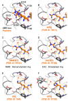
The first pocket of the UBR box recognizes the first residue of type 1 N-degrons. (A) Residues constituting the first pocket have enough space (black dotted line) for positively charged residues to enter. (BF) Hydrogen bonding and charge–charge interactions (yellow dotted lines) generated when positively charged residues and modified arginine residues bind to the pockets, and water molecules (red dots) fill the empty spaces.
Figure 5
Figure 5
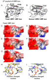
The second pocket of the UBR box recognizes the second residue of type 1 N-degron. (A,B) Residues constituting the second pocket of the UBR boxes of yeast UBR1 and human UBR2 are represented by sticks (PDB ID 3NIH and 3NY3). (C,D) These surface models interact with hydrophobic residues (Ile and Leu). Blue on the surface means a positively charged, and red means a negatively charged. The ligands are bound to the hydrophobic surface of this pocket (PDB ID 3NIH and 3NIN). (E,F) Acidic residues (Asp and Glu) are bound to the UBR box. T171 is pushed back by the negatively charged of acidic residues (PDB ID 3NIL and 3NIK). (G) The basic residue Arg is bound to the UBR box. It can be seen that R135 is pulled back to bind the large and positively charged Arg residue (PDB ID 3NIM). (H,I) The changes in the secondary pocket that occur when charged residues bind are represented by cartoon and stick models (PDB ID 3NIM and 3NIK). (J,K) The ligand structures bound to the human UBR box are shown as surface and stick models with a larger surface than yeast because residues such as R135 and T171 do not exist in the human UBR box (PDB ID 3NY3 and 5TDA). (L) The secondary pocket of the human UBR box and its ligands are shown as cartoon and stick model (PDB ID 3NY3 and 5TDA).
Figure 5
Figure 5
The second pocket of the UBR box recognizes the second residue of type 1 N-degron. (A,B) Residues constituting the second pocket of the UBR boxes of yeast UBR1 and human UBR2 are represented by sticks (PDB ID 3NIH and 3NY3). (C,D) These surface models interact with hydrophobic residues (Ile and Leu). Blue on the surface means a positively charged, and red means a negatively charged. The ligands are bound to the hydrophobic surface of this pocket (PDB ID 3NIH and 3NIN). (E,F) Acidic residues (Asp and Glu) are bound to the UBR box. T171 is pushed back by the negatively charged of acidic residues (PDB ID 3NIL and 3NIK). (G) The basic residue Arg is bound to the UBR box. It can be seen that R135 is pulled back to bind the large and positively charged Arg residue (PDB ID 3NIM). (H,I) The changes in the secondary pocket that occur when charged residues bind are represented by cartoon and stick models (PDB ID 3NIM and 3NIK). (J,K) The ligand structures bound to the human UBR box are shown as surface and stick models with a larger surface than yeast because residues such as R135 and T171 do not exist in the human UBR box (PDB ID 3NY3 and 5TDA). (L) The secondary pocket of the human UBR box and its ligands are shown as cartoon and stick model (PDB ID 3NY3 and 5TDA).
Figure 6
Figure 6
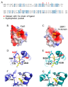
Homology models of the human UBR1 N-domain based on ClpS of Escherichia coli. The homology model of the N-domain of human UBR1 was generated based on the ClpS structure (PDB ID 2W9R) using the SWISS-MODEL program [65,66]. (A) The amino acid sequences of human UBR1 and E. coli ClpS are aligned. Conserved residues are highlighted with light blue boxes. Red and light green dots indicate the residues recognizing type 2 N-degrons. (B,C) The surface models of the ClpS and N-domain are colored by charge. The ligand, Phe–Arg peptide, is represented as a stick. (D,E) The residues constituting the entrance of the hydrophobic pocket interact with the backbone of the first amino acid (Phe) by charge–charge interactions and hydrogen bonding (red dotted lines). (F,G) The hydrophobic residue of the first amino acid is bound to the hydrophobic pocket. The red dotted lines mark the space created by the hydrophobic residues constituting the pocket.
Figure 7
Figure 7
A model describing the regulatory role of UBR1 and UBR2 in G-protein signaling pathways via mediating RGS protein degradation. In GPCRs (G-protein-coupled receptors), heterotrimeric G-proteins are dissociated into G〈 and G®© subunits by external ligands or signal mediators. Activated GTP-bound G〈 and G®© stimulate the downstream signaling pathway associated with cell growth and cardiovascular development. The proper regulation of GTP-Gα activity by GTPase-activating RGS proteins is vital in the GPCR-related signaling pathway. Among the RGS proteins, RGS4, RGS5, and RGS16 are cleaved by MetAP to expose a cysteine residue at the N-terminus. After which, these RGS proteins undergo oxidation, followed by arginylation (C* denotes the oxidized N-terminal Cysteine residue). Arginylated RGS proteins are recognized by UBR1 and UBR2 for ubiquitination and degradation. When the Arg/N-degron pathway is genetically inhibited, metabolically stabilized RGS proteins promote the hydrolysis of the GTP-bounded G〈 subunit, which leads to inactivation of the GPCR signaling pathway.
Figure 8
Figure 8
A model depicting the degradation of pro-apoptotic fragments mediated by UBR1/2 in response to apoptotic stimuli. Caspases or calpains generate pro-apoptotic fragments during the induction of apoptosis. These proteins expose destabilizing residues at the N-terminus, which are short-lived N-degron substrates. As a result, these pro-apoptotic fragments are selectively degraded by the Arg/N-degron pathway, contributing to cell survival. As a negative feedback mechanism, caspases can also inhibit UBR1 function by inducing its cleavage.
Figure 9
Figure 9
A schematic model describing mitochondrial quality control mediated by UBR1/2/4 in response to the mitochondrial damage. (A) PINK1 is continuously imported into healthy mitochondria through TOM and TIM complexes under steady-state conditions. After which, the precursor PINK1 is cleaved by MPP and PARL proteases, respectively. PARL cleaves between Ala103 and Phe104 of PINK1 to expose phenylalanine, a known type-2 destabilizing residue of the Arg/N-degron pathway, at the N-terminus. The cleaved PINK1 is released into the cytosol. Phe104-PINK is recognized by UBR1, UBR2, and UBR4 for ubiquitination and proteasomal degradation. (B) When depolarization or mitochondrial import is blocked, PINK1 accumulates on the OMM. The accumulation and autophosphorylation of PINK1 recruit Parkin E3 ligase from the cytosol to the mitochondria, leading to the degradation of damaged mitochondria through mitophagy.
Figure 10
Figure 10
Schematic diagrams describing the regulation of inflammation by UBR box N-recognins. (A) A model depicting LT-induced mNLRPB inflammasome activation by UBR2/4. The anthrax lethal factor (LF) is known to induce mNLRP1B inflammasome activation and pyroptosis. Although the detailed mechanism of LT-induced mNLRP1B inflammasome activation has not been elucidated, it has recently been shown that the Arg/N-degron pathway is involved in mNLRP1B inflammasome activation. LF directly cleaves mNLRP1B to generate an N-terminal fragment and a C-terminal fragment. Despite cleavage into N-terminal and C-terminal fragments by LF, mNLRP1B remains autoinhibited. To activate the inflammasome, the N-terminal fragment of mNLRP1B is degraded in an Arg/N-degron pathway-dependent manner by UBR2 and UBR4, as identified through CRISPR-Cas9 screening. This process releases the CARD domain-containing C-terminal fragment of mNLRP1B and induces pyroptosis through interaction with caspase-1. (B) A model depicting the degradation of proinflammatory fragments mediated by UBR1/2/4/5. Potential proinflammatory Arg/N-degron substrates are generated by activated inflammatory caspases and several other proteases under inflammatory stimuli such as LPS. The resulting N-degron substrates Cys-RAB39a, Asn-CASP1, Gln-CASP4, CASP5, Ile-GRZA, and Ile-GRZM are generated by activated caspases, autoprocessing, or by endopeptidases such as DPP1. These fragments, which can cause an inflammatory response, expose destabilizing residues at the N-terminus. These fragments are recognized by N-recognins and then degraded through 26S proteasome, as evidenced by the depletion of UBR1, UBR2, UBR4, and UBR5, causing a significant reduction in LPS-induced IL-1β secretion.
Figure 11
Figure 11
UBR1/2 mediated SDE2’s C-terminal fragment degradation upon replication stress, leading to recovering a stalled replication fork. The irradiation of cells with ultraviolet C, one of the causes of DNA replication stress, results in DNA lesions that block replication. The Arg/N-degron pathway is involved in counteracting DNA replication stress. Monoubiquitination of PCNA plays a vital role in coordinating DNA repair against replication-blocked lesions by providing a platform to recruit factors necessary for DNA repair. To counteract DNA replication stress, the C-terminal fragment of SDE2 needs to be degraded (A). Under UV-induced replication stress, SDE2 is targeted to the replication fork by interacting with PCNA through the PIP box of the UBL domain. After which, a C-terminal fragment of SDE2 (SDECt) is generated by the cleavage of the diglycine motif by DUB. Intriguingly, SDECt inhibits UV damage-inducible PCNA monoubiquitination by RAD18 ubiquitin E3 ligase. (B) Damage-inducible SDE2 Ct phosphorylation of Ser266 or Thr319, or both by ATR. (C) SDE2Ct, which has an N-terminal lysine, is recognized by UBR1 and UBR2 for polyubiquitination. In addition, phosphorylated SDE2Ct facilitates the interaction of p97UFD1-NPL4 and enables the extraction of ubiquitinated SDE2Ct from chromatin. (D) Consequently, degradation of SDECt by the Arg/N-degron pathway-ATR- p97UFD1-NPL4 axis promotes the monoubiquitination of PCNA by RAD18 E3 ligase, leading to DNA damage bypass, stalled fork recovery, and S phase progression.

References

    1. Dikic I. Proteasomal and Autophagic Degradation Systems. Annu. Rev. Biochem. 2017;86:193–224. doi: 10.1146/annurev-biochem-061516-044908. - DOI - PubMed
    1. Zhao J., Zhai B., Gygi S.P., Goldberg A.L. mTOR inhibition activates overall protein degradation by the ubiquitin proteasome system as well as by autophagy. Proc. Natl. Acad. Sci. USA. 2015;112:15790–15797. doi: 10.1073/pnas.1521919112. - DOI - PMC - PubMed
    1. King B., Trimarchi T., Reavie L., Xu L., Mullenders J., Ntziachristos P., Aranda-Orgilles B., Perez-Garcia A., Shi J., Vakoc C., et al. The ubiquitin ligase FBXW7 modulates leukemia-initiating cell activity by regulating MYC stability. Cell. 2013;153:1552–1566. doi: 10.1016/j.cell.2013.05.041. - DOI - PMC - PubMed
    1. Zolk O., Schenke C., Sarikas A. The ubiquitin-proteasome system: Focus on the heart. Cardiovasc. Res. 2006;70:410–421. doi: 10.1016/j.cardiores.2005.12.021. - DOI - PubMed
    1. Rahimi N. The ubiquitin-proteasome system meets angiogenesis. Mol. Cancer Ther. 2012;11:538–548. doi: 10.1158/1535-7163.MCT-11-0555. - DOI - PMC - PubMed

Substances

LinkOut - more resources