. 2021 Aug 3;22(15):8336.
doi: 10.3390/ijms22158336.

Inflammasome Signaling Regulates the Microbial-Neuroimmune Axis and Visceral Pain in Mice

Affiliations

Inflammasome Signaling Regulates the Microbial-Neuroimmune Axis and Visceral Pain in Mice

Mònica Aguilera et al. Int J Mol Sci. .

Abstract

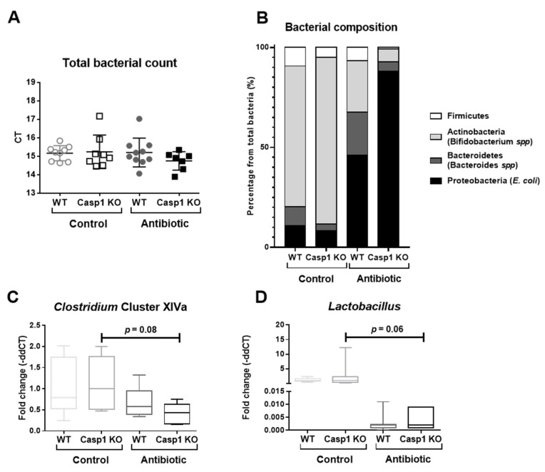
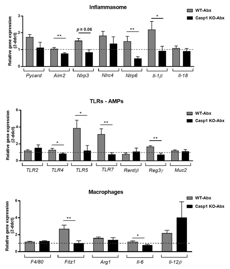
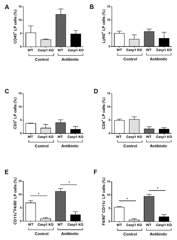
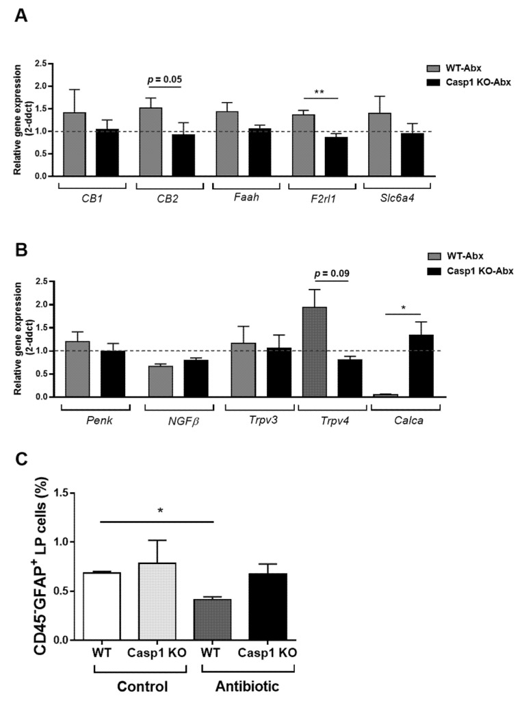
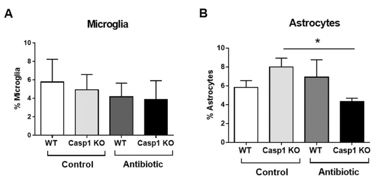
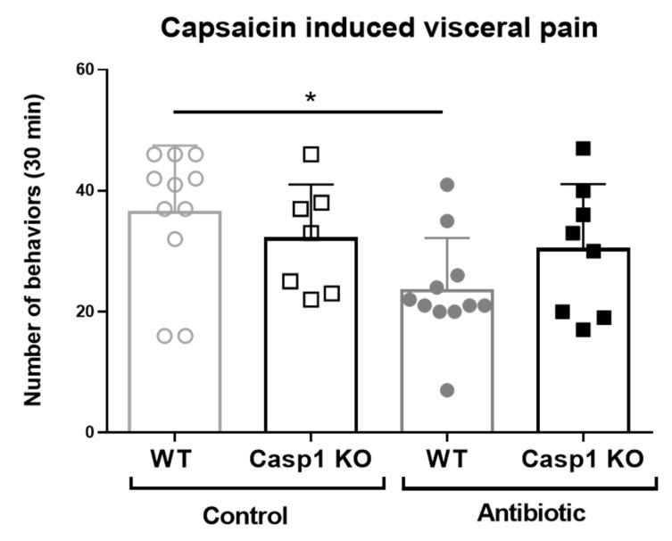
Interactions between the intestinal microbiota, immune system and nervous system are essential for homeostasis in the gut. Inflammasomes contribute to innate immunity and brain-gut interactions, but their role in microbiota-neuro-immune interactions is not clear. Therefore, we investigated the effect of the inflammasome on visceral pain and local and systemic neuroimmune responses after antibiotic-induced changes to the microbiota. Wild-type (WT) and caspase-1/11 deficient (Casp1 KO) mice were orally treated for 2 weeks with an antibiotic cocktail (Abx, Bacitracin A and Neomycin), followed by quantification of representative fecal commensals (by qPCR), cecal short chain fatty acids (by HPLC), pathways implicated in the gut-neuro-immune axis (by RT-qPCR, immunofluorescence staining, and flow cytometry) in addition to capsaicin-induced visceral pain responses. Abx-treatment in WT-mice resulted in an increase in colonic macrophages, central neuro-immune interactions, colonic inflammasome and nociceptive receptor gene expression and a reduction in capsaicin-induced visceral pain. In contrast, these responses were attenuated in Abx-treated Casp1 KO mice. Collectively, the data indicate an important role for the inflammasome pathway in functional and inflammatory gastrointestinal conditions where pain and alterations in microbiota composition are prominent.

Keywords: gut commensal microbiota; gut–brain axis; immune system; inflammasome.


Conflict of interest statement

FS is the co-founder/shareholder of Alimentary Health Ltd., Tucana Health Ltd. and Atlantia Food Clinical Trials Ltd. and scientific advisor to Kaleido Biosciences. All other authors declare no conflict of interest. The funders had no role in the design of the study; in the collection, analyses, or interpretation of data; in the writing of the manuscript, or in the decision to publish the results.

Figures

Figure 1
Figure 1
Characterization and quantification of total bacteria, the phylum Firmicutes, Actinobacteria, Bacteroidetes and Proteobacteria, and the genera Clostridium XIVa and Lactobacillus by qPCR in control and antibiotic-treated WT and Casp1 KO mice. (A) Representative cycle threshold (CT) values for total bacterial qPCR detection. (B) Percentage (%) of the composition including Firmicutes, Actinobacteria (Bifidobacterium spp), Bacteroidetes (Bacteroides spp) and Proteobacteria (E. coli), representing the commensal microbiota from the total bacterial abundance and (C) abundance of Clostridium cluster XIVa and (D) Lactobacillus Order of the Firmicutes phylum. (A) Data are presented as mean (SD), n = 7–10/group. (B) Data are presented as percentage of each phylum from the fold change of the total bacterial abundance. (C,D) Data are median (interquartile range) (SD).
Figure 2
Figure 2
Representative RT-qPCR expression of genes associated with (A) inflammasome (Pycard, Aim2, Nlrp3, Nlrc4, Nlrp6, Il-1β and Il-18); (B) toll-like receptors (TLR2, -4, -5 and -7); antimicrobial peptides (AMPs-Rentlβ and Reg3γ), the mucus layer component Muc2 and (C) macrophage M1/M2 signature (F4/80, Fitz1, Arg1, Il-6 and Il-12β), the colon of WT and Casp1 KO antibiotic-treated groups. Data are mean (SEM). n= 7–9/group. *: p < 0.05, **: p <0.001. Dashed line indicates a background value of 1 of WT and Casp1 KO-control groups. Abx: Antibiotic.
Figure 3
Figure 3
Colonic lamina propria (LP) cells isolated from control and antibiotic-treated WT and Casp1 KO mice and stained with (A) CD45 (Leukocytes); (B) Ly6G (neutrophils); (C) CD3 (CD3+ T cells); (D) CD4 (CD4+ T cells); (E) CD11c (dendritic cells); and (F) F4/80 (macrophages). Bars represent the percentage of the indicated cell population. Representative figure of two individual experiments. Data are mean (SEM). n = 4–5/group. *: p < 0.05.
Figure 4
Figure 4
Representative RT-qPCR expression of colon nociceptive markers; (A) the endocannabinoid system (CB1, CB2 and Faah), the protease-activated receptor 2 (PAR2, Fr2l1) and the serotonin transporter (Scl6a4); (B) the opioid peptide pro-enkephalin (Penk), the neurothrophin (NGFβ), the vanilloid system (transient receptor potential, Trpv3 and Trpv4) and calcitonin-related polypeptide alpha (Calca). Data are mean (SEM). n = 7–9/group. *: p <0.05, **: p < 0.001. Abx: antibiotic. Dashed line indicates background value of 1 of WT and Casp1 KO-control groups. (C) Colonic lamina propria (LP) cells isolated from control and antibiotic-treated WT and Casp1 KO mice and stained with CD45 and GFAP. Bars represent the percentage of the indicated cell population. Representative figure of two individual experiments. Data are mean (SD). n = 4/group. *: p < 0.05.
Figure 5
Figure 5
Representative percentage (%) of (A) microglia and (B) astrocytes cell average in the anterior cingulate cortex (ACC) of WT and Casp1 KO control and antibiotic-treated groups. Data are mean (SD). n = 3/group. *: p < 0.05.
Figure 6
Figure 6
Representative number of visceral pain behaviors during 30 min per each experimental group upon intracolonic administration of capsaicin. Data mean (SD), * p < 0.05 compared to the WT control group. n = 7–11/group, including in all groups, males (range 2 to 8) and females (range 3 to 5).

Similar articles

Cited by

References

    1. Statovci D., Aguilera M., MacSharry J., Melgar S. The Impact of Western Diet and Nutrients on the Microbiota and Immune Response at Mucosal Interfaces. Front. Immunol. 2017;8:838. doi: 10.3389/fimmu.2017.00838. - DOI - PMC - PubMed
    1. Chen Y., Wu G., Zhao Y. Gut Microbiota and Alimentary Tract Injury. Adv. Exp. Med. Biol. 2020;1238:11–22. doi: 10.1007/978-981-15-2385-4_2. - DOI - PubMed
    1. Man S.M. Inflammasomes in the gastrointestinal tract: Infection, cancer and gut microbiota homeostasis. Nat. Rev. Gastroenterol. Hepatol. 2018;15:721–737. doi: 10.1038/s41575-018-0054-1. - DOI - PMC - PubMed
    1. Black C.J., Drossman D.A., Talley N.J., Ruddy J., Ford A.C. Functional gastrointestinal disorders: Advances in understanding and management. Lancet. 2020;396:1664–1674. doi: 10.1016/S0140-6736(20)32115-2. - DOI - PubMed
    1. Zhen Y., Zhang H. NLRP3 Inflammasome and Inflammatory Bowel Disease. Front. Immunol. 2019;10:276. doi: 10.3389/fimmu.2019.00276. - DOI - PMC - PubMed

MeSH terms