Review
. 2021 Jul 26;14(15):4151.
doi: 10.3390/ma14154151.

Advanced Optogenetic-Based Biosensing and Related Biomaterials

Affiliations
Review

Advanced Optogenetic-Based Biosensing and Related Biomaterials

Mihaela Gheorghiu et al. Materials (Basel). .

Abstract

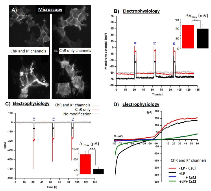
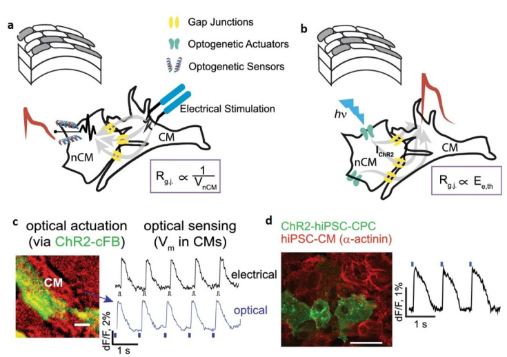
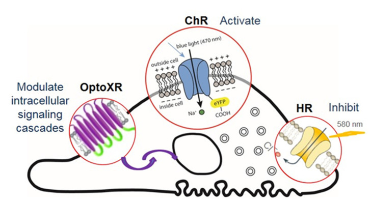
The ability to stimulate mammalian cells with light, brought along by optogenetic control, has significantly broadened our understanding of electrically excitable tissues. Backed by advanced (bio)materials, it has recently paved the way towards novel biosensing concepts supporting bio-analytics applications transversal to the main biomedical stream. The advancements concerning enabling biomaterials and related novel biosensing concepts involving optogenetics are reviewed with particular focus on the use of engineered cells for cell-based sensing platforms and the available toolbox (from mere actuators and reporters to novel multifunctional opto-chemogenetic tools) for optogenetic-enabled real-time cellular diagnostics and biosensor development. The key advantages of these modified cell-based biosensors concern both significantly faster (minutes instead of hours) and higher sensitivity detection of low concentrations of bioactive/toxic analytes (below the threshold concentrations in classical cellular sensors) as well as improved standardization as warranted by unified analytic platforms. These novel multimodal functional electro-optical label-free assays are reviewed among the key elements for optogenetic-based biosensing standardization. This focused review is a potential guide for materials researchers interested in biosensing based on light-responsive biomaterials and related analytic tools.

Keywords: cell dynamics; cell-based biosensors; optogenetic (light-responsive) biomaterials; optogenetic control; time-lapse multiparametric assays.


Conflict of interest statement

The authors declare no conflict of interest.

Figures

Scheme 1
Scheme 1
An overview of membrane-acting optogenetic control, and compatible phenotyping readouts across relevant cell-based biosensing platforms, from single cells to 3D tissue mimics (organoids), to control and assess the dynamics of cellular responses.
Figure 1
Figure 1
Complementary characterization of the functional expression of genetically encoded ion channels (the ChR2 and ROMK1 channels). Microscopy (A): different levels of ChR2-YFP expression can be quantified using fluorescence imaging (fluorescence images, scale bar 10 µm) as well as their membrane distribution using Total internal Fluorescence Microscopy (lower panel scale bar 1 µm). Electrophysiology: (B) Current clamp data reveal the (resting) membrane potential values (Vm) and their changes upon illumination as a function of channel repertoire; inset membrane potential values changes (ΔVmax = Vmax (pulse) − Vm) upon illumination—enabling comparison between light-induced depolarization changes (significance level ** p < 0.01, n =5) for ChR-only expression and ChR and K+ channels, respectively. (C) The Voltage clamp data reveal transmembrane currents (I) upon illumination and enable quantification of functional channel density. Inset, the amplitude (ΔImax = I0− Imax (pulse)) of the current elicited by the first LP is significantly (*** p < 0.001, n = 5) higher for cells expressing ChR only, corresponding to the functional ChR2 channels opening. For cells with a more complex channel repertoire (e.g., cells expressing both ChR and K+ channels), separating the individual transmembrane current components require specific channel inhibitors (see D). (D) Representative I-V plots corresponding to −80 mV ÷ +60 mV voltage ramps with K+ channel inhibitor CsCl and on–off illumination (green/blue curves) demonstrating functional, tandem expression of K+ channels and ChR2 channels (as controls we use −LP-CsCl as well as +LP-CsCl current values, i.e., red and black tracks, respectively).
Scheme 2
Scheme 2
Optogenetic methods offer new ways for assessing hetero-cellular coupling as either (a) ‘optogenetic–sensor’ variant, where coupling is typically confirmed by measuring membrane potential fluctuations (VnCM) in non-cardiomyocytes (nCMs) cells expressing genetically encoded voltage indicator (GEVI)/ genetically encoded calcium indicator (GECI) and connected to cardiomyocytes CMs undergoing excitation or (b) ‘optogenetic–actuator’ approach where coupling can be quantified by the light needed to trigger excitation in the CMs via the light-sensitive non-cardiomyocyte nCMs type cells, i.e., Ee,th. This ‘optogenetic–actuator’ approach is the recommended way to implement the tool for both ChR2-cFB and CMs (c) as well as for light-sensitized human cardiac progenitor cells (ChR2-iPS-CPC) and non-transformed human induced pluripotent stem cells derived CMs (hiPSC-CMs) (d). Redrawn from [54].
Scheme 3
Scheme 3
(a) Confocal of compacted toroid neural tissue mimic (NTM). (Scale bar: 1 mm.) (b) Installation of toroid NTMs (Day 4 from seeding) on functionalized glass cylinders after compaction in the 3D-printed mold. (c) LightSheet image of neurites extending throughout the glass rod (β-TubulinIII, green; F-actin, yellow) after 3 days. (Scale bar: 1 mm; 500 μm.) Redrawn from ([37], PNAS 2019].
Figure 2
Figure 2
Common light-sensitive proteins in optogenetics research: channelrhodopsin ChR allows inward photocurrent of non-selective cations (Na+ > K+ >> H+ >> Ca2+), which can evoke cell depolarization under 470 nm wavelength light (with activating effect for excitable cells). Halorhodopsin HR, an anion (Cl) channel/pump, can produce membrane hyperpolarization and electrophysiological inhibition under 580 nm wavelength light. OptoXR (a G-protein-coupled receptor) is used to modulate intracellular signaling cascades.
Figure 3
Figure 3
Schematic representation of a cell-based impedance sensing system with optogenetic control. The cells grown on the electrode act as insulators, impeding the flow of current, perturbing the impedance of the system as revealed by multi frequency Electrical Impedance Spectroscopy (EIS). Upon light control of optogenetic actuators, the characteristic impedance response (black) is rapidly and specifically modified upon addition of compounds able to alter individual cell characteristics (cell morphology, cytoskeleton, metabolism, cell signaling) or to disrupt the cell monolayer (affecting cell–cell communication or cell-surface attachment). Access to the whole dynamics, including the recovery phase, has predictive potential concerning the persistence of the compound-specific bio-effect (i.e., altered homeostasis/homeostasis restoration).

References

    1. Li Y.E., Lee I.C. The Current Trends of Biosensors in Tissue Engineering. Biosensors. 2020;10:88. doi: 10.3390/bios10080088. - DOI - PMC - PubMed
    1. Lee J.H., Luo J., Choi H.K., Chueng ST D., Lee K.B., Choi J.W. Functional nanoarrays for investigating stem cell fate and function. Nanoscale. 2020;12:9306–9326. doi: 10.1039/C9NR10963C. - DOI - PMC - PubMed
    1. Gheorghiu M. A short review on cell-based biosensing: Challenges and breakthroughs in biomedical analysis. J. Biomed Res. 2020:1–9. doi: 10.7555/jbr.34.20200128. - DOI - PMC - PubMed
    1. Gupta N., Renugopalakrishnan V., Liepmann D., Paulmurugan R., Malhotra B.D. Cell-based biosensors: Recent trends, challenges and future perspectives. Biosens. Bioelectron. 2019;141:111435. doi: 10.1016/j.bios.2019.111435. - DOI - PubMed
    1. Xie M., Fussenegger M. Designing cell function: Assembly of synthetic gene circuits for cell biology applications. Nat. Rev. Mol. Cell Biol. 2018;19:507–525. doi: 10.1038/s41580-018-0024-z. - DOI - PubMed

LinkOut - more resources