. 2021 Jul 21;10(15):3198.
doi: 10.3390/jcm10153198.

An Assistive Role of a Machine Learning Network in Diagnosis of Middle Ear Diseases

Affiliations

An Assistive Role of a Machine Learning Network in Diagnosis of Middle Ear Diseases

Hayoung Byun et al. J Clin Med. .

Abstract

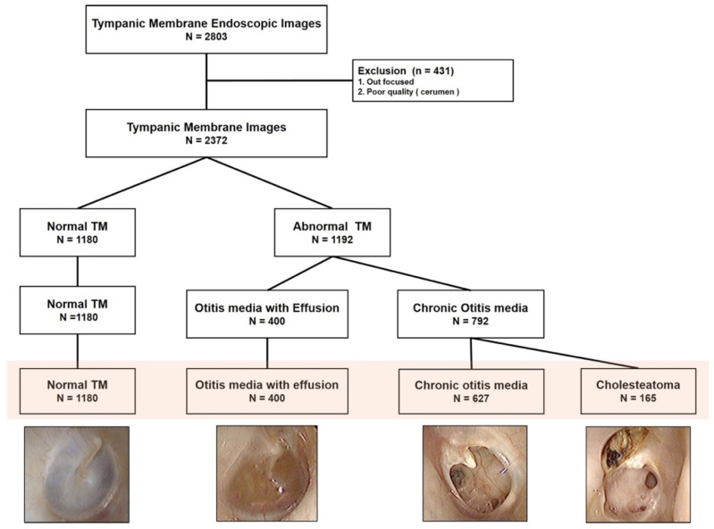
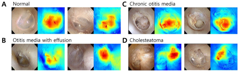
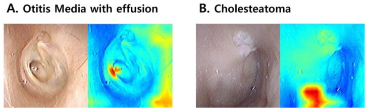
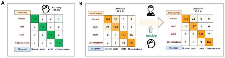
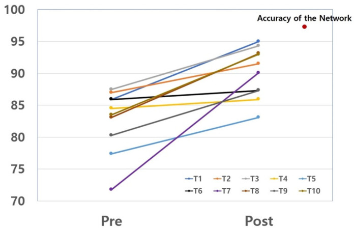
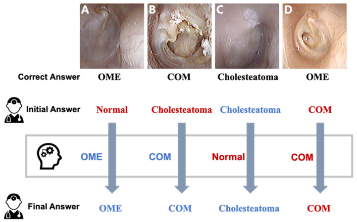
The present study aimed to develop a machine learning network to diagnose middle ear diseases with tympanic membrane images and to identify its assistive role in the diagnostic process. The medical records of subjects who underwent ear endoscopy tests were reviewed. From these records, 2272 diagnostic tympanic membranes images were appropriately labeled as normal, otitis media with effusion (OME), chronic otitis media (COM), or cholesteatoma and were used for training. We developed the "ResNet18 + Shuffle" network and validated the model performance. Seventy-one representative cases were selected to test the final accuracy of the network and resident physicians. We asked 10 resident physicians to make diagnoses from tympanic membrane images with and without the help of the machine learning network, and the change of the diagnostic performance of resident physicians with the aid of the answers from the machine learning network was assessed. The devised network showed a highest accuracy of 97.18%. A five-fold validation showed that the network successfully diagnosed ear diseases with an accuracy greater than 93%. All resident physicians were able to diagnose middle ear diseases more accurately with the help of the machine learning network. The increase in diagnostic accuracy was up to 18% (1.4% to 18.4%). The machine learning network successfully classified middle ear diseases and was assistive to clinicians in the interpretation of tympanic membrane images.

Keywords: artificial intelligence; machine learning; otitis media; resident physician; tympanic membrane.


Conflict of interest statement

The authors declare no conflict of interest.

Figures

Figure 1
Figure 1
Acquisition and labeling of the tympanic membrane images.
Figure 2
Figure 2
Network architecture for tympanic membrane diagnosis.
Figure 3
Figure 3
The regions of interest for tympanic membrane diagnosis were visualized as a heat map using Gradient-weighted Class Activation Mapping (Grad-CAM). (A) Grad-CAM focused on the pars tensa region of the normal tympanic membrane. (B) The change in color of the tympanic membrane due to middle ear effusion is important for the diagnosis of otitis media with effusion in the Grad-CAM analysis. (C) Chronic otitis media could be distinguished by perforation and remnant tympanic membrane. (D) In cholesteatoma, the location of the bony erosion was spotted in Grad-CAM.
Figure 4
Figure 4
Gradient-weighted Class Activation Mapping (Grad-CAM) of the incorrectly predicted tympanic membrane images. (A) Otitis media with effusion was misdiagnosed as chronic otitis media because the machine learning model focused on the retracted part of the tympanic membrane and considered it as a perforation. (B) The machine learning model predicted a normal tympanic membrane by focusing on the pars tensa area of the tympanic membrane. The heat map image failed to highlight the small attic retraction and cholesteatoma materials.
Figure 5
Figure 5
The confusion matrix for the diagnostic results for the machine learning network and resident physicians. (A) Prediction accuracy of the machine learning network in the representative cases, (B) The diagnostic performance change of ten resident physicians with the assistance of the machine learning network.
Figure 6
Figure 6
The diagnostic accuracy for middle ear diseases was increased in resident physicians after consultation with the machine learning model.
Figure 7
Figure 7
Representative cases in tympanic membrane images. (A,B) Many resident physicians changed to the correct answer with the advice of the machine learning network. (C) All resident physicians diagnosed cholesteatoma correctly despite wrong advice. (D) Resident physicians and the machine learning misdiagnosed OME as COM. OME: otitis media with effusion. COM: Chronic otitis media.

References

    1. Miller D.D., Brown E.W. Artificial Intelligence in Medical Practice: The Question to the Answer? Am. J. Med. 2018;131:129–133. doi: 10.1016/j.amjmed.2017.10.035. - DOI - PubMed
    1. Chassagnon G., Vakalopoulou M., Paragios N., Revel M.P. Artificial intelligence applications for thoracic imaging. Eur. J. Radiol. 2020;123:108774. doi: 10.1016/j.ejrad.2019.108774. - DOI - PubMed
    1. Jeon Y., Lee K., Sunwoo L., Choi D., Oh D.Y., Lee K.J., Kim Y., Kim J.W., Cho S.J., Baik S.H., et al. Deep Learning for Diagnosis of Paranasal Sinusitis Using Multi-View Radiographs. Diagnostics. 2021;11:250. doi: 10.3390/diagnostics11020250. - DOI - PMC - PubMed
    1. Xu J., Wang S., Zhou Z., Liu J., Jiang X., Chen X. Automatic CT image segmentation of maxillary sinus based on VGG network and improved V-Net. Int. J. Comput. Assist. Radiol. Surg. 2020;15:1457–1465. doi: 10.1007/s11548-020-02228-6. - DOI - PubMed
    1. Lee J. Is Artificial Intelligence Better Than Human Clinicians in Predicting Patient Outcomes? J. Med. Internet Res. 2020;22:e19918. doi: 10.2196/19918. - DOI - PMC - PubMed

LinkOut - more resources