. 2021 Jul 21;10(15):3206.
doi: 10.3390/jcm10153206.

Ultrasound Patterns in the First Trimester Diagnosis of Congenital Heart Disease

Affiliations

Ultrasound Patterns in the First Trimester Diagnosis of Congenital Heart Disease

Catalin G Herghelegiu et al. J Clin Med. .

Abstract

Congenital heart disease (CHD) is the most common birth defect, with a reported prevalence of 5-12 per 1000 live births. Very recently, the American Institute of Ultrasound in Medicine published a guideline recommending the use of the four-chamber and the three-vessel and trachea views to screen for CHD in the first trimester of pregnancy. Our aim is to present abnormal image patterns that are seen in the four-chamber, three-vessel, and trachea views of the fetal heart in the first trimester and to describe their association with specific CHD types. We used a total of 29 cases of CHD from the archives of Filantropia Hospital and the Maternal and Child Health Institute (INSMC) fetal medicine units. We selected cases with a clear and well-documented diagnosis of the CHD type. We identified a series of repeating color doppler flow patterns seen in the four-chamber, three-vessel, and trachea views of the studied cases. Our observations could be developed into a diagnosis algorithm to orientate the examiner to the most likely type of CHD in individual cases.

Keywords: congenital heart disease; first trimester of pregnancy; four-chamber view; three-vessel and trachea views.


Conflict of interest statement

The authors declare no conflict of interest.

Figures

Figure 1
Figure 1
Color doppler flow patterns encountered in the four-chamber view. (A): Normal filling of both ventricles. (B): Filling of both ventricles, but the cardiac axis is abnormally shifted to the left. (C): Common inflow into the ventricles. (D): Filling of both ventricles, but the left ventricle appears smaller. (E): Filling of only one ventricle.
Figure 2
Figure 2
Color doppler flow patterns encountered in the three-vessel and trachea view. (A): Typical V sign with equally sized arms. (B): The vessels forming a U sign around the trachea. (C): V sign with a narrower arm. (D): One vessel with an oblique and curved course. (E): One vessel with a straight course.
Figure 3
Figure 3
Color doppler flow patterns seen in the three-vessel and trachea view in the case of CAT. (A): Pulmonary arteries arising from the vessel. (B): The straight course of the vessel through the mediastinum.
Figure 4
Figure 4
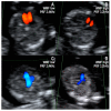
Color doppler flow patterns of the four-chamber, and three-vessel, and trachea views obtained using low and high PRF/WMF doppler settings in a normal 12–13-week fetus. (A): Low PRF/WMF doppler settings allow better visualization of the size and filling of the ventricles in the four-chamber view. (B): High PRF/WMF doppler settings are preferred to verify the distinct filling of the ventricles. (C): Low PRF/WMF doppler settings offer adequate visualization of the ductal and aortic arches. (D): High PRF/WMF doppler settings can impair the visualization of the aorta.

References

    1. Donofrio M.T., Moon-Grady A.J., Hornberger L.K., Copel J.A., Sklansky M.S., Abuhamad A., Cuneo B.F., Huhta J.C., Jonas R.A., Krishnan A., et al. Diagnosis and Treatment of Fetal Cardiac Disease. Circulation. 2014;129:2183–2242. doi: 10.1161/01.cir.0000437597.44550.5d. - DOI - PubMed
    1. Hoffman J.I.E., Kaplan S. The incidence of congenital heart disease. J. Am. Coll. Cardiol. 2002;39:1890–1900. doi: 10.1016/S0735-1097(02)01886-7. - DOI - PubMed
    1. Becker R., Wegner R.D. Detailed screening for fetal anomalies and cardiac defects at the 11–13-week scan. Ultrasound Obstet. Gynecol. 2006;27:613–618. doi: 10.1002/uog.2709. - DOI - PubMed
    1. Rasiah S.V., Publicover M., Ewer A.K., Khan K.S., Kilby M.D., Zamora J. A systematic review of the accuracy of first-trimester ultrasound examination for detecting major congenital heart disease. Ultrasound Obstet. Gynecol. 2006;28:110–116. doi: 10.1002/uog.2803. - DOI - PubMed
    1. Smrcek J.M., Berg C., Geipel A., Fimmers R., Axt-Fliedner R., Diedrich K., Gembruch U. Detection rate of early fetal echocardiography and in utero development of congenital heart defects. J. Ultrasound Med. 2006;25:187–196. doi: 10.7863/jum.2006.25.2.187. - DOI - PubMed

LinkOut - more resources