Review
. 2021 Jul 22;10(15):3227.
doi: 10.3390/jcm10153227.

Pulmonary Complications in Hematopoietic Stem Cell Transplant Recipients-A Clinician Primer

Affiliations
Review

Pulmonary Complications in Hematopoietic Stem Cell Transplant Recipients-A Clinician Primer

Anna Astashchanka et al. J Clin Med. .

Abstract

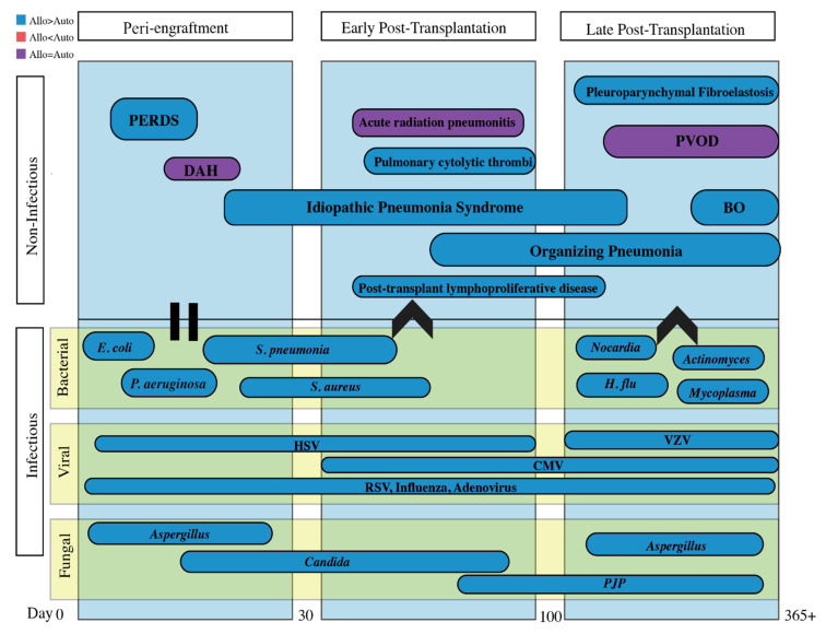
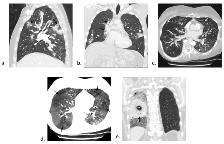
Hematopoietic stem cell transplants (HSCT) are becoming more widespread as a result of optimization of conditioning regimens and prevention of short-term complications with prophylactic antibiotics and antifungals. However, pulmonary complications post-HSCT remain a leading cause of morbidity and mortality and are a challenge to clinicians in both diagnosis and treatment. This comprehensive review provides a primer for non-pulmonary healthcare providers, synthesizing the current evidence behind common infectious and non-infectious post-transplant pulmonary complications based on time (peri-engraftment, early post-transplantation, and late post-transplantation). Utilizing the combination of timing of presentation, clinical symptoms, histopathology, and radiographic findings should increase rates of early diagnosis, treatment, and prognostication of these severe illness states.

Keywords: DAH; IPS; PERDS; bronchiolitis obliterans syndrome; post-HSCT.


Conflict of interest statement

The authors declare no conflict of interest.

Figures

Figure 1
Figure 1
A timetable organized by peri-engraftment, early post-transplantation and late post-transplantation periods encompassing both infectious and non-infectious pulmonary complications. Important to note that all of the above vary in time of presentation and can often overlap. For example, the increase in non-myeloablative conditioning can make this timeline less reliable. Infectious and non-infectious complications tend to occur more commonly in allogeneic transplants are typically secondary to chronic graft-versus-host disease and prolonged immunosuppressive medications. Abbreviations: PERDS = peri-engraftment respiratory distress syndrome; DAH = diffuse alveolar hemorrhage; PVOD = pulmonary veno-occlusive disease; BO = bronchiolitis obliterans; HSV = herpes simplex virus; VZV = varicella zoster virus; RSV = Respiratory syncytial virus; CMV = cytomegalovirus; PJP = Pneumocystis jirovecii.
Figure 2
Figure 2
CT findings of peri-engraftment non-infectious pulmonary complications. a–c: Peri-engraftment respiratory distress syndrome (PERDS) in a 28-year-old woman with a fever and rash who is 7 days post-autologous HSCT for acute myelogenous leukemia (AML). (a) Axial image from a chest CT shows diffuse ground-glass opacity with interlobular and intralobular septal thickening creating a “crazy paving” pattern (black arrow). This is a nonspecific finding that can be seen in PERDS, DAH, viral pneumonia, and pulmonary edema. At the time of this scan, there was no lower lobe volume loss (white arrow). Despite steroid therapy, the patient’s symptoms worsened. (b) Axial image from a chest CT four days later shows increased lower lobe ground-glass opacity and septal thickening. The left major fissure is posteriorly displaced (white arrow) due to increasing lower lobe volume loss. (c) Axial image from a chest CT 11 days later shows findings of acute respiratory distress syndrome (ARDS) with extensive lower lobe predominant consolidation and ground-glass opacity with bronchial dilation (black arrow) and increasing lower lobe volume loss (white arrow). The patient developed pneumomediastinum (asterisk) due to barotrauma from intubation. (d) Diffuse alveolar hemorrhage (DAH) in a 32-year-old man 14 days status post-autologous HSCT. Axial CT image shows lower lobe predominant ground-glass opacity with a small focus of consolidation at the left base. Diffuse interlobular and intralobular septal thickening creating a “crazy paving” pattern (black arrow) is present. While this pattern has a broad differential diagnosis, DAH was confirmed on bronchoscopy.
Figure 3
Figure 3
CT findings in early-post transplant non-infectious complications. (a) Axial CT in a 25-year-old man 89-days post-autologous HSCT for aplastic anemia shows lower lobe predominant consolidation. After an extensive work-up, the patient was diagnosed with idiopathic pneumonia syndrome (IPS). He died 24 days later. (b) Axial chest CT shows numerous enlarged right axillary lymph nodes (white arrow). (c) Coronal image from a subsequent positron emission tomography (PET)-CT shows increased FDG uptake in the right axillary nodes as well as uptake in right cervical and mesenteric lymph nodes (white arrows) consistent with a diagnosis of PTLD.
Figure 4
Figure 4
CT findings in late non-infectious complications. (a,b) Bronchiolitis obliterans syndrome (BOS) in a 42-year old woman two years after autologous HSCT. (a) Axial imaging during inspiration demonstrates a subtle mosaic attenuation with multiple areas of increased lucency (asterisks) adjacent to more normal lung. (b) On expiratory scan, the normal lung increases in attenuation. However, the areas with BO do not change in attenuation (asterisks) on expiration due to air trapping. (c) Chronic graft-versus-host disease (cGVHD) manifesting as organizing pneumonia (OP) in a 53-year-old man 5 months after autologous HSCT. Axial CT shows lower lobe and subpleural predominant consolidation and ground-glass opacity (black arrows) with a few scattered nodules. An “atoll” sign is present in the right lower lobe (white arrow). Extensive infectious work-up was negative and open lung biopsy showed organizing pneumonia. (df) Pleuroparenchymal fibroelastosis (PPFE) and bronchiolitis obliterans syndrome (BOS) in a 36-year-old woman 21 years after autologous HSCT for myelodysplastic syndrome. (d) Posteroanterior (PA) radiograph shows pronounced pleural thickening superiorly (black arrows) with superior displacement of the hila and upper lobe bronchiectasis (white arrows). (e) Corresponding coronal image from a chest CT better shows the exuberant superior pleural thickening extending inferior along the periphery of the upper lobes (black arrows). There is extensive upper lobe volume loss with superior displacement of the hila and areas of conglomerate perihilar and upper lobe fibrosis (white arrow). A mosaic attenuation is present in the lower lobes with areas of relative increased attenuation (asterisks). (f) A 10 mm thick minimum intensity projection image (MinIP) better shows the upper lobe predominant bronchiectasis (white arrows) and the areas of relative hypoattenuation in the lower lobes (asterisks). The patient underwent lung transplant which confirmed both PPFE and BO.
Figure 5
Figure 5
CT findings in early and late infectious complications. (a)Angioinvasive aspergillus in a 37-year-old woman status post-autologous HSCT for acute myeloid leukemia (AML). Sagittal chest CT shows numerous areas of nodular consolidation with surrounding ground-glass halos (black arrows) consistent with an angioinvasive infection. Aspergillus was confirmed on bronchoscopy. (b) Streptococcus pneumoniae infection in a 34-year-old man status post-autologous HSCT for Hodgkin lymphoma. Coronal chest CT shows nonspecific areas of consolidation in the left upper lobe and right lower lobe (white arrows). Streptococcus pneumoniae infection was confirmed on bronchoscopy. Paramediastinal fibrosis in the right lung (black arrows) due to radiation therapy for HL can be seen. (c,d) Nontuberculous mycobacterial infection (NTM) and bronchiolitis obliterans syndrome (BOS) in a 51-year-old woman three years after autologous stem cell transplant for acute lymphoblastic leukemia. (c) A 5 mm thick maximum intensity projection image (MIP) shows bronchial wall thickening and multifocal tree-in-bud nodularity (white arrows) due to NTM infection. (d) Corresponding 5 mm thick minimum intensity projection image (MinIP) at the same level nicely shows the pronounced mosaic attenuation (black arrows) due to BOS. (e) Mucor infection in a 49-year-old man nine months after autologous HSCT. Coronal image from a chest CT shows a mass-like lesion with dense circular consolidation (black arrow) with central necrosis manifesting as “bubbly”-appearing areas of ground-glass opacity (white arrow) and cavitation (asterisk). This “bird’s nest” sign is highly suggesting of mucormycosis. Surrounding ground-glass opacity with a “crazy paving” pattern (black arrowhead) is due to hemorrhage.

Similar articles

Cited by

References

    1. Barriga F., Ramirez P., Wietstruck A., Rojas N. Hematopoietic stem cell transplantation: Clinical use and perspectives. Biol. Res. 2012;45:307–316. doi: 10.4067/S0716-97602012000300012. - DOI - PubMed
    1. Cordonnier C., Bernaudin J.F., Bierling P., Huet Y., Vernant J.P. Pulmonary complications occurring after allogeneic bone marrow transplantation. A study of 130 consecutive transplanted patients. Cancer. 1986;58:1047–1054. doi: 10.1002/1097-0142(19860901)58:5<1047::AID-CNCR2820580512>3.0.CO;2-Y. - DOI - PubMed
    1. Jules-Elysee K., Stover D.E., Yahalom J., White D.A., Gulati S.C. Pulmonary complications in lymphoma patients treated with high-dose therapy autologous bone marrow transplantation. Am. Rev. Respir Dis. 1992;146:485–491. doi: 10.1164/ajrccm/146.2.485. - DOI - PubMed
    1. Shorr A.F., Moores L.K., Edenfield W.J., Christie R.J., Fitzpatrick T.M. Mechanical ventilation in hematopoietic stem cell transplantation: Can We effectively predict outcomes? Chest. 1999;116:1012–1018. doi: 10.1378/chest.116.4.1012. - DOI - PubMed
    1. Forman S.J., Negrin R.S., Antin J.H., Appelbaum F.R. Thomas’ Hematopoietic Cell Transplantation. Wiley Blackwell; Hoboken, NJ, USA: 2016.