

Biology and Management of Dedifferentiated Liposarcoma: State of the Art and Perspectives
- PMID: 34362013
- PMCID: PMC8348700
- DOI: 10.3390/jcm10153230
Biology and Management of Dedifferentiated Liposarcoma: State of the Art and Perspectives
Abstract
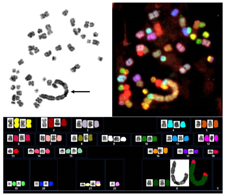
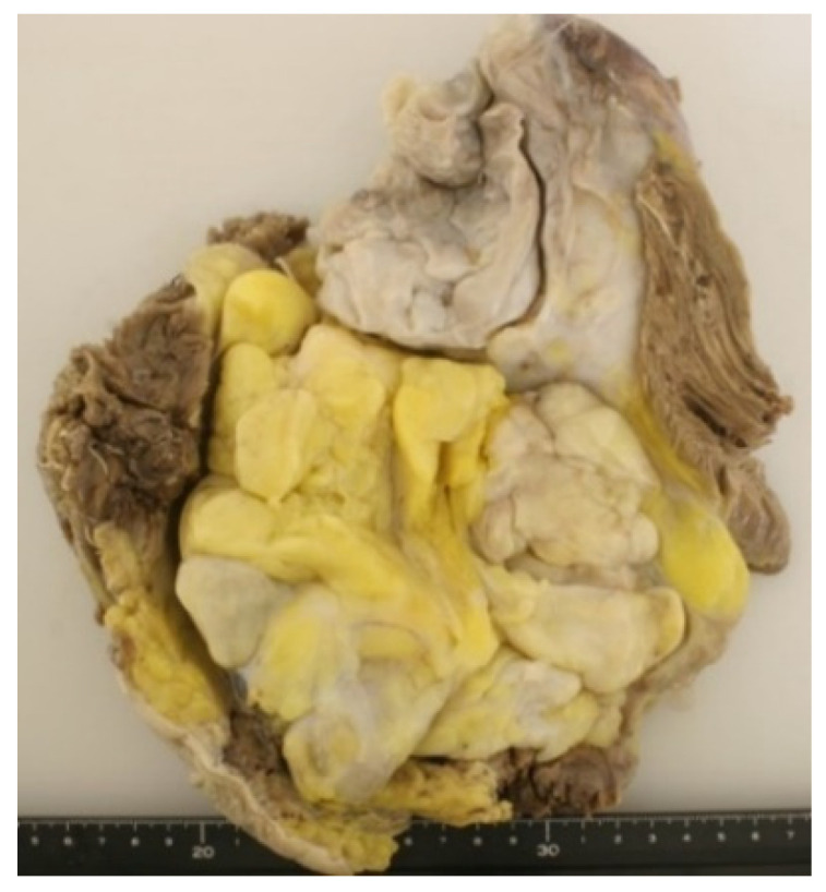
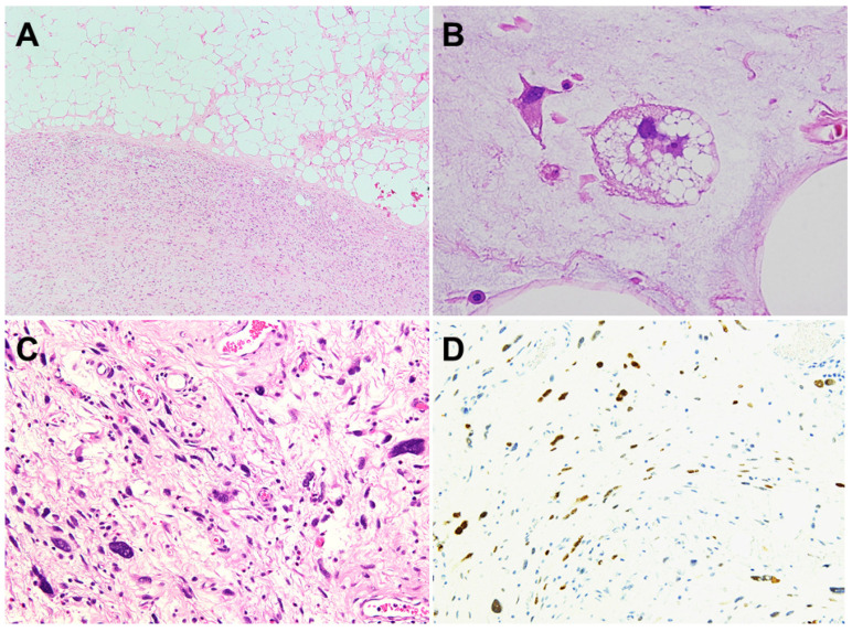
Dedifferentiated liposarcoma (DDL) is defined as the transition from well-differentiated liposarcoma (WDL)/atypical lipomatous tumor (ALT) to non-lipogenic sarcoma, which arises mostly in the retroperitoneum and deep soft tissue of proximal extremities. It is characterized by a supernumerary ring and giant marker chromosomes, both of which contain amplified sequences of 12q13-15 including murinedouble minute 2 (MDM2) and cyclin-dependent kinase 4 (CDK4) cell cycle oncogenes. Detection of MDM2 (and/or CDK4) amplification serves to distinguish DDL from other undifferentiated sarcomas. Recently, CTDSP1/2-DNM3OS fusion genes have been identified in a subset of DDL. However, the genetic events associated with dedifferentiation of WDL/ALT remain to be clarified. The standard treatment for localized DDL is surgery, with or without radiotherapy. In advanced disease, the standard first-line therapy is an anthracycline-based regimen, with either single-agent anthracycline or anthracycline in combination with the alkylating agent ifosfamide. Unfortunately, this regimen has not necessarily led to a satisfactory clinical outcome. Recent advances in the understanding of the pathogenesis of DDL may allow for the development of more-effective innovative therapeutic strategies. This review provides an overview of the current knowledge on the clinical presentation, pathogenesis, histopathology and treatment of DDL.
Keywords: atypical lipomatous tumor; dedifferentiated liposarcoma; diagnosis; pathogenesis; treatment; well-differentiated liposarcoma.
Conflict of interest statement
The authors declare no conflict of interest.
Figures
Similar articles
-
Well-differentiated liposarcoma and dedifferentiated liposarcoma: An updated review.Semin Diagn Pathol. 2019 Mar;36(2):112-121. doi: 10.1053/j.semdp.2019.02.006. Epub 2019 Feb 28. Semin Diagn Pathol. 2019. PMID: 30852045 Review.
-
Expression of FRS2 in atypical lipomatous tumor/well-differentiated liposarcoma and dedifferentiated liposarcoma: an immunohistochemical analysis of 182 cases with genetic data.Diagn Pathol. 2021 Oct 25;16(1):96. doi: 10.1186/s13000-021-01161-9. Diagn Pathol. 2021. PMID: 34696768 Free PMC article.
-
Overlapping features between dedifferentiated liposarcoma and undifferentiated high-grade pleomorphic sarcoma.Am J Surg Pathol. 2009 Nov;33(11):1594-600. doi: 10.1097/PAS.0b013e3181accb01. Am J Surg Pathol. 2009. PMID: 19574885
-
A novel sclerosing atypical lipomatous tumor/well-differentiated liposarcoma in a 7-year-old girl: report of a case with molecular confirmation.Hum Pathol. 2018 Jan;71:41-46. doi: 10.1016/j.humpath.2017.06.015. Epub 2017 Jul 11. Hum Pathol. 2018. PMID: 28705709
-
Dedifferentiated Liposarcoma: Updates on Morphology, Genetics, and Therapeutic Strategies.Adv Anat Pathol. 2016 Jan;23(1):30-40. doi: 10.1097/PAP.0000000000000101. Adv Anat Pathol. 2016. PMID: 26645460 Review.
Cited by
-
Atypical Spindle Cell/Pleomorphic Lipomatous Tumor: A Review and Update.Cancers (Basel). 2024 Sep 13;16(18):3146. doi: 10.3390/cancers16183146. Cancers (Basel). 2024. PMID: 39335118 Free PMC article. Review.
-
Beyond targeting amplified MDM2 and CDK4 in well differentiated and dedifferentiated liposarcomas: From promise and clinical applications towards identification of progression drivers.Front Oncol. 2022 Sep 2;12:965261. doi: 10.3389/fonc.2022.965261. eCollection 2022. Front Oncol. 2022. PMID: 36119484 Free PMC article. Review.
-
Giant dedifferentiated liposarcoma of the gastrocolic ligament: A case report.World J Gastrointest Surg. 2023 Oct 27;15(10):2376-2381. doi: 10.4240/wjgs.v15.i10.2376. World J Gastrointest Surg. 2023. PMID: 37969706 Free PMC article.
-
Recurrent dedifferentiated liposarcoma with histological grade progression: a case report.Ecancermedicalscience. 2025 Jan 22;19:1831. doi: 10.3332/ecancer.2025.1831. eCollection 2025. Ecancermedicalscience. 2025. PMID: 40177153 Free PMC article.
-
The pyroptosis-related signature predicts prognosis and influences the tumor immune microenvironment in dedifferentiated liposarcoma.Open Med (Wars). 2024 Jan 9;19(1):20230886. doi: 10.1515/med-2023-0886. eCollection 2024. Open Med (Wars). 2024. PMID: 38221934 Free PMC article.
References
-
- The WHO Classification of Tumors Editorial Board . WHO Classification of Tumours of Soft Tissue and Bone. 5th ed. IARC Press; Lyon, France: 2020. pp. 36–48.
-
- Waters R., Horvai A., Greipp P., John I., Demicco E.G., Dickson B.C., Tanas M., Larsen B.T., Din N.U., Creytens D.H., et al. Atypical lipomatous tumour/well-differentiated liposarcoma and de-differentiated liposarcoma in patients aged ≤40 years: A study of 116 patients. Histopathology. 2019;75:833–842. doi: 10.1111/his.13957. - DOI - PubMed
Publication types
Grants and funding
LinkOut - more resources
Full Text Sources
Research Materials
