

Retinal OCT Findings in Patients after COVID Infection
- PMID: 34362017
- PMCID: PMC8347407
- DOI: 10.3390/jcm10153233
Retinal OCT Findings in Patients after COVID Infection
Abstract
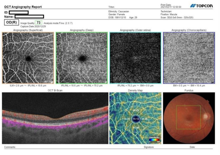
Purpose: The aim of this study was to assess and compare the optic nerve, retina, and retinal vessel parameters in recovered COVID-19 patients and healthy patients by using optical coherence tomography angiography (OCT-a).
Methods: In all, 156 eyes of post-COVID-19 patients and 98 eyes of subjects from a control group were enrolled in our study. BCVA, intra ocular pressure (IOP) measurement, fundus examination, and OCT images, including macular cube, OCT-RNFL, and angio-OCT 6 × 6 mm examinations, were performed for both groups. The measurements were acquired using Swept Source OCT DRI OCT Triton. In the post-COVID-19 group, 762 OCT protocols were obtained. For statistical analysis, parameters from only one eye from each subject were taken.
Results: In the measured parameters, no significant differences were observed, i.e., central macular thickness (p = 0.249); RNFL (p = 0.104); FAZ (p = 0.63); and vessel density of superficial retinal vascular plexus in central (p = 0.799), superior (p = 0.767), inferior (p = 0.526), nasal (p = 0.402), and temporal (p = 0.582) quadrants. Furthermore, a slit-lamp examination did not reveal any COVID-19-related abnormalities.
Conclusion: OCT examination did not detect any significant changes in morphology or morphometry of the optic nerve, retina, or the retina vessels due to COVID-19.
Keywords: COVID-19; angiography; choroid; optical coherence tomography; retina; vascular complications.
Conflict of interest statement
The authors declare no conflict of interest.
Figures
Similar articles
-
Optical coherence tomography of retinal and choroidal layers in patients with familial hypercholesterolaemia treated with lipoprotein apheresis.Atheroscler Suppl. 2019 Dec;40:49-54. doi: 10.1016/j.atherosclerosissup.2019.08.031. Atheroscler Suppl. 2019. PMID: 31818450
-
Evaluation of the effect of energy drink consumption on retina and choroid: an optical coherence tomography and optical coherence tomography angiography study.Cutan Ocul Toxicol. 2020 Dec;39(4):295-297. doi: 10.1080/15569527.2020.1755977. Epub 2020 Sep 14. Cutan Ocul Toxicol. 2020. PMID: 32285710 Clinical Trial.
-
[Correlation of capillary plexus with visual acuity in idiopathic macular epiretinal membrane eyes using optical coherence tomography angiography].Zhonghua Yan Ke Za Zhi. 2019 Oct 11;55(10):757-762. doi: 10.3760/cma.j.issn.0412-4081.2019.10.006. Zhonghua Yan Ke Za Zhi. 2019. PMID: 31607064 Chinese.
-
Retinal and Choroidal Changes in Patients with Parkinson's Disease Detected by Swept-Source Optical Coherence Tomography.Curr Eye Res. 2018 Jan;43(1):109-115. doi: 10.1080/02713683.2017.1370116. Epub 2017 Nov 7. Curr Eye Res. 2018. PMID: 29111842
-
Humphrey matrix frequency doubling technology perimetry and optical coherence tomography measurement of the retinal nerve fiber layer thickness in both normal and ocular hypertensive subjects.J Glaucoma. 2006 Aug;15(4):328-35. doi: 10.1097/01.ijg.0000212230.65545.d3. J Glaucoma. 2006. PMID: 16865011
Cited by
-
The Ocular Surface Symptoms and Tear Film Parameters during and after COVID-19 Infection.J Clin Med. 2022 Nov 12;11(22):6697. doi: 10.3390/jcm11226697. J Clin Med. 2022. PMID: 36431174 Free PMC article.
-
Reduced Vessel Density and Enlarged Foveal Avascular Zone in the Macula as a Result of Systemic Hypoxia Caused by SARS-CoV-2 Infection.J Pers Med. 2023 May 31;13(6):926. doi: 10.3390/jpm13060926. J Pers Med. 2023. PMID: 37373915 Free PMC article. Review.
-
Retinal Microvascular Changes in COVID-19 Bilateral Pneumonia Based on Optical Coherence Tomography Angiography.J Clin Med. 2022 Jun 23;11(13):3621. doi: 10.3390/jcm11133621. J Clin Med. 2022. PMID: 35806907 Free PMC article.
-
Long-Term Effects of COVID-19 on Optic Disc and Retinal Microvasculature Assessed by Optical Coherence Tomography Angiography.Diagnostics (Basel). 2025 Jan 6;15(1):114. doi: 10.3390/diagnostics15010114. Diagnostics (Basel). 2025. PMID: 39795642 Free PMC article.
-
Headache and cognitive disturbance correlate with ganglion cell layer thickness in patients who recovered from COVID-19.Clin Neurol Neurosurg. 2022 Jun;217:107263. doi: 10.1016/j.clineuro.2022.107263. Epub 2022 Apr 26. Clin Neurol Neurosurg. 2022. PMID: 35525105 Free PMC article.
References
-
- Soiza R.L., Donaldson A.I.C., Myint P.K. Vaccine against arteriosclerosis: An update. Ther. Adv. Vaccines. 2018;9:259–261.
-
- Abrishami M., Emamverdian Z., Shoeibi N., Omidtabrizi A., Daneshvar R., Rezvani T.S., Saeedian N., Eslami S., Mazloumi M., Sadda S., et al. Optical coherence tomography angiography analysis of the retina in patients recovered from COVID-19: A case-control study. Can. J. Ophthalmol. 2021;56:24–30. doi: 10.1016/j.jcjo.2020.11.006. - DOI - PMC - PubMed
LinkOut - more resources
Full Text Sources
