Visual Acuity and Number of Amniotic Membrane Layers as Indicators of Efficacy in Amniotic Membrane Transplantation for Corneal Ulcers: A Multicenter Study
- PMID: 34362018
- PMCID: PMC8348886
- DOI: 10.3390/jcm10153234
Visual Acuity and Number of Amniotic Membrane Layers as Indicators of Efficacy in Amniotic Membrane Transplantation for Corneal Ulcers: A Multicenter Study
Abstract
Background: To evaluate new indicators in the efficacy of amniotic membrane transplantation (AMT) for non-healing corneal ulcers (NHCUs).
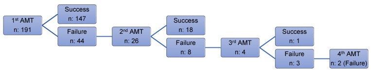
Methods: Retrospective, multicenter study. In total, 223 AMTs for NHCU in 191 patients were assessed. The main outcomes studied were the success rate of AMT (complete re-epithelization), postoperative visual acuity (VA) gain, and number of AM layers transplanted.
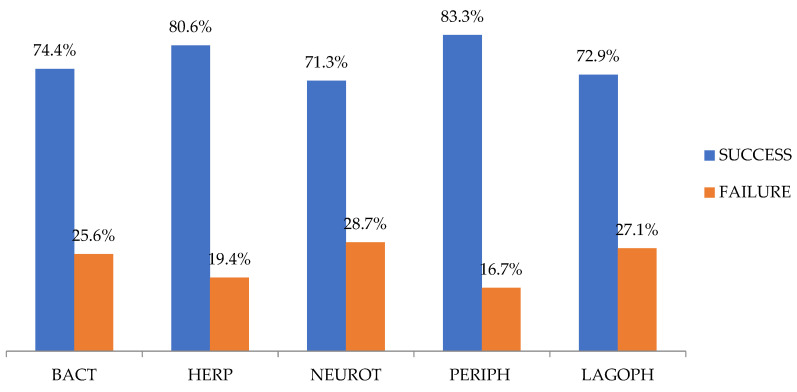
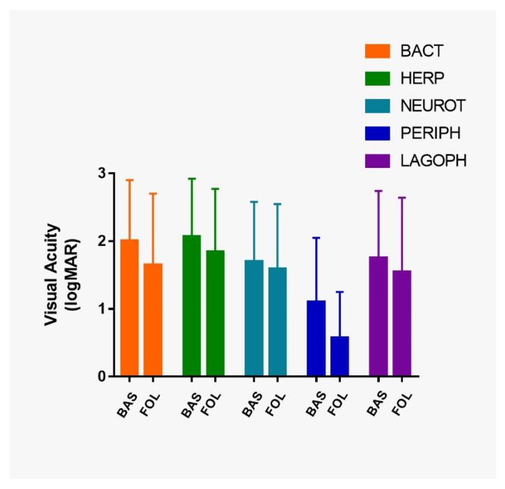
Results: The overall AMT success rate was 74.4%. In 92% of our patients VA stability or improvement. Postoperative VA was significantly higher than preoperative VA in the entire cohort (p < 0.001) and in all etiological groups of ulcers (post-bacterial, p ≤ 0.001; post-herpetic, p ≤ 0.0038; neurotrophic ulcers, p ≤ 0.014; non-rheumatic peripheral, p ≤ 0.001; and ulcers secondary to lagophthalmos and eyelid malposition or trauma, p ≤ 0.004). Most participants (56.5%) presented a preoperative VA equal to or less than counting fingers (≤0.01). Of these, 13.5% reached a postoperative VA equal to or better than legal blindness (≥0.05) after AMT. A higher success rate was observed in the monolayer than in the multilayer AMT (79.5% and 64.9%, respectively; p = 0.018). No statistically significant values were found between the number of layers transplanted and VA gain (p = 0.509).
Conclusion: AMT is not only beneficial in achieving complete re-epithelialization in NHCUs but also in improving postoperative VA; these improvements are independent of etiologies of ulcers. Furthermore, the use of monolayer AMT seems to be a more appropriate option than multilayer AMT for NHCU since the multilayer AMT did not present better outcomes (success rate and VA gain) compared to monolayer AMT in the different types of ulcers studied.
Keywords: amniotic membrane; amniotic membrane transplantation; cornea; corneal ulcer; corneal ulceration; non healing corneal ulcer; persistent epithelial defects; visual acuity.
Conflict of interest statement
The authors have no conflict of interest to declare.
Figures