

Comparative analyses of two primate species diverged by more than 60 million years show different rates but similar distribution of genome-wide UV repair events
- PMID: 34362292
- PMCID: PMC8349011
- DOI: 10.1186/s12864-021-07898-3
Comparative analyses of two primate species diverged by more than 60 million years show different rates but similar distribution of genome-wide UV repair events
Abstract
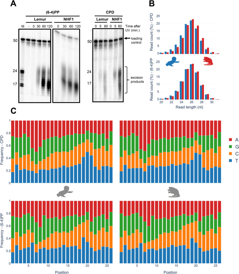
Background: Nucleotide excision repair is the primary DNA repair mechanism that removes bulky DNA adducts such as UV-induced pyrimidine dimers. Correspondingly, genome-wide mapping of nucleotide excision repair with eXcision Repair sequencing (XR-seq), provides comprehensive profiling of DNA damage repair. A number of XR-seq experiments at a variety of conditions for different damage types revealed heterogenous repair in the human genome. Although human repair profiles were extensively studied, how repair maps vary between primates is yet to be investigated. Here, we characterized the genome-wide UV-induced damage repair in gray mouse lemur, Microcebus murinus, in comparison to human.
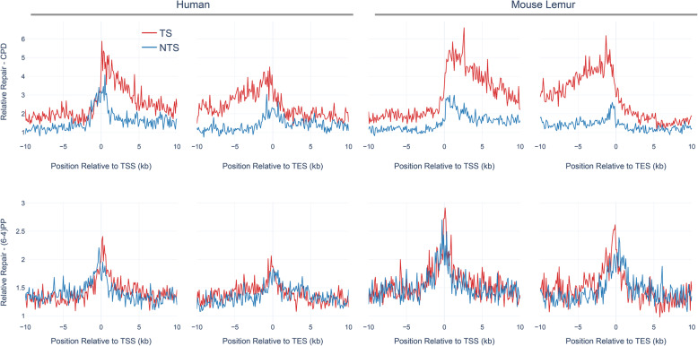
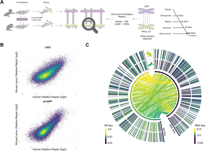
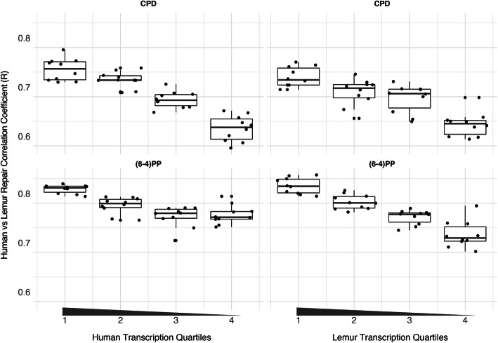
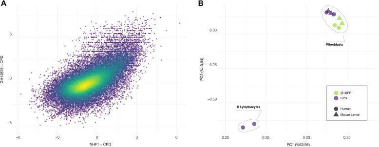
Results: We derived fibroblast cell lines from mouse lemur, exposed them to UV irradiation, and analyzed the repair events genome-wide using the XR-seq protocol. Mouse lemur repair profiles were analyzed in comparison to the equivalent human fibroblast datasets. We found that overall UV sensitivity, repair efficiency, and transcription-coupled repair levels differ between the two primates. Despite this, comparative analysis of human and mouse lemur fibroblasts revealed that genome-wide repair profiles of the homologous regions are highly correlated, and this correlation is stronger for highly expressed genes. With the inclusion of an additional XR-seq sample derived from another human cell line in the analysis, we found that fibroblasts of the two primates repair UV-induced DNA lesions in a more similar pattern than two distinct human cell lines do.
Conclusion: Our results suggest that mouse lemurs and humans, and possibly primates in general, share a homologous repair mechanism as well as genomic variance distribution, albeit with their variable repair efficiency. This result also emphasizes the deep homologies of individual tissue types across the eukaryotic phylogeny.
Keywords: (6–4)PP; CPD; Mouse Lemur; Nucleotide excision repair; Primate; UV damage; XR-seq.
© 2021. The Author(s).
Conflict of interest statement
The authors declare that there is no competing interests regarding the publication of this article.
Figures

References
-
- Hu J, Selby CP, Adar S, Adebali O, Sancar A. Molecular mechanisms and genomic maps of DNA excision repair in Escherichia coli and humans. J Biol Chem. 2017; https://doi.org/gftwfh. 10.1074/jbc.r117.807453. PMID: 28798238. PMCID: PMC5612094. - PMC - PubMed
-
- Li W, Sancar A. Methodologies for detecting environmentally induced DNA damage and repair. Environ Mol Mutagen. 2020; https://doi.org/ggmr42. 10.1002/em.22365. PMID: 32083352. PMCID: PMC7442611. - PMC - PubMed
-
- Hu J, Adebali O, Adar S, Sancar A. Dynamic maps of UV damage formation and repair for the human genome. Proc Natl Acad Sci. 2017; https://doi.org/ggdfws. 10.1073/pnas.1706522114. PMID: 28607063. PMCID: PMC5495279. - PMC - PubMed
-
- Mao P, Brown AJ, Esaki S, Lockwood S, Poon GMK, Smerdon MJ, et al. ETS transcription factors induce a unique UV damage signature that drives recurrent mutagenesis in melanoma. Nat Commun. 2018; https://doi.org/gdxcvk. 10.1038/s41467-018-05064-0. PMID: 29980679. PMCID: PMC6035183. - PMC - PubMed
-
- Hu J, Adar S, Selby CP, Lieb JD, Sancar A. Genome-wide analysis of human global and transcription-coupled excision repair of UV damage at single-nucleotide resolution. Genes Dev. 2015; https://doi.org/f69xk2. 10.1101/gad.261271.115. PMID: 25934506. PMCID: PMC4421983. - PMC - PubMed
MeSH terms
Substances
Grants and funding
LinkOut - more resources
Full Text Sources
Molecular Biology Databases
Miscellaneous
