. 2021 Aug 6;14(1):123.
doi: 10.1186/s13041-021-00836-0.

Inhibition of autophagy by CRMP2-derived peptide ST2-104 (R9-CBD3) via a CaMKKβ/AMPK/mTOR pathway contributes to ischemic postconditioning-induced neuroprotection against cerebral ischemia-reperfusion injury

Affiliations

Inhibition of autophagy by CRMP2-derived peptide ST2-104 (R9-CBD3) via a CaMKKβ/AMPK/mTOR pathway contributes to ischemic postconditioning-induced neuroprotection against cerebral ischemia-reperfusion injury

Yuan Yao et al. Mol Brain. .

Abstract

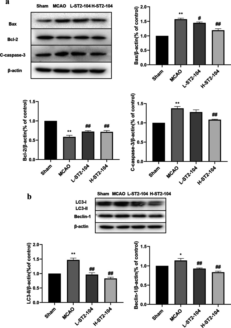
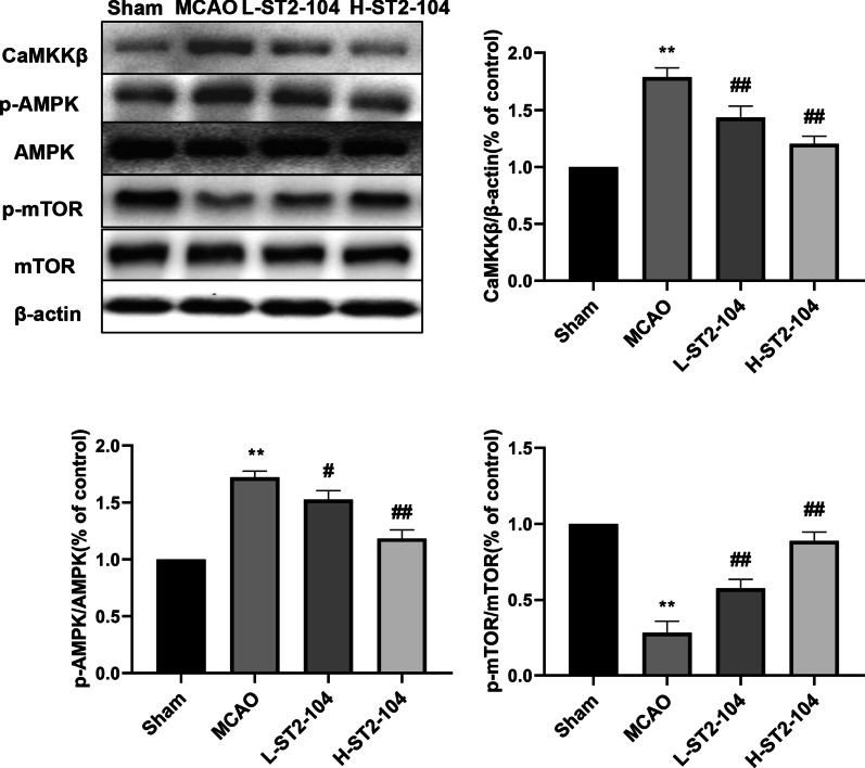
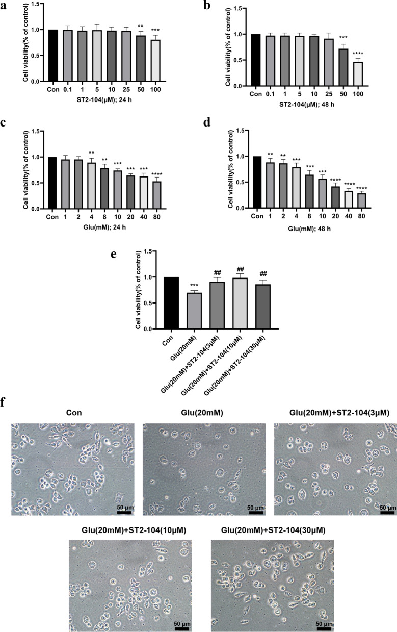
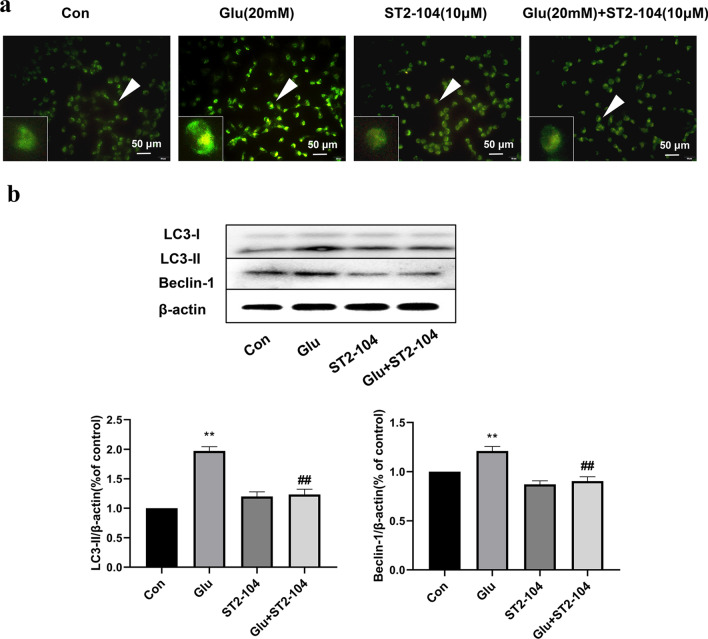
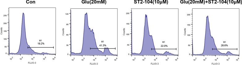
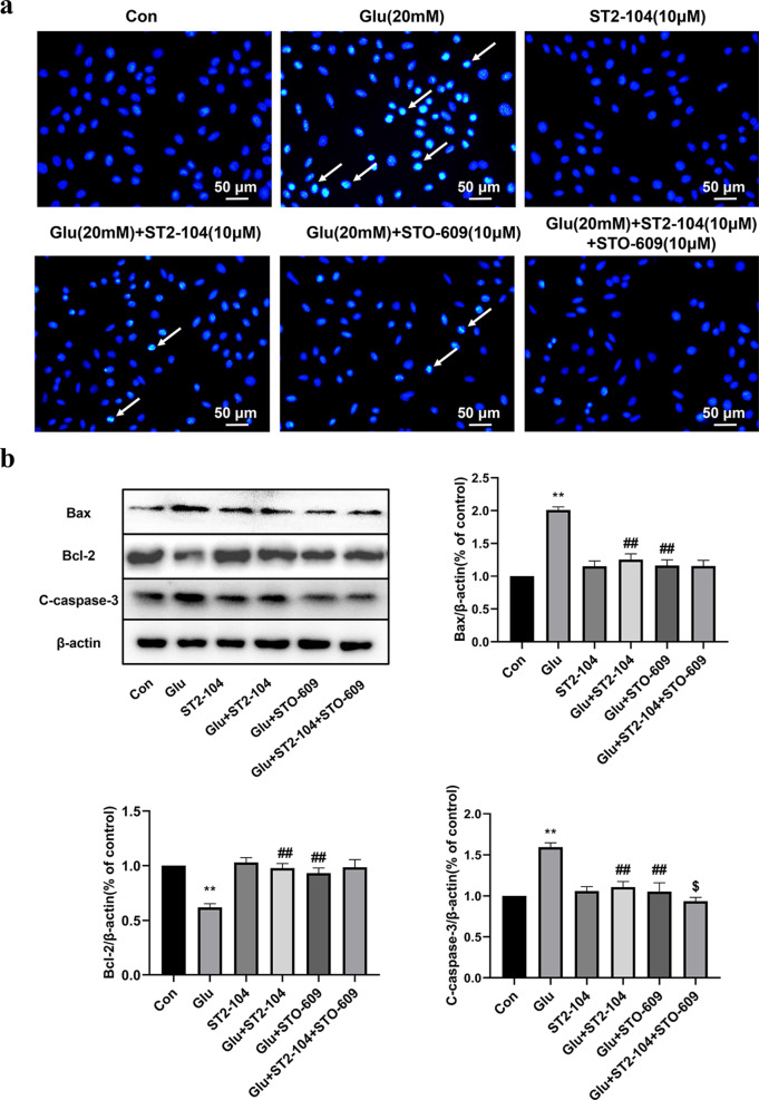
Cerebral ischemia, a common cerebrovascular disease, is characterized by functional deficits and apoptotic cell death. Autophagy, a type of programmed cell death, plays critical roles in controlling neuronal damage and metabolic homeostasis, and has been inextricably linked to cerebral ischemia. We previously identified a short peptide aptamer from collapsin response mediator protein 2 (CRMP2), designated the Ca2+ channel-binding domain 3 (CBD3) peptide, that conferred protection against excitotoxicity and traumatic brain injury. ST2-104, a nona-arginine (R9)-fused CBD3 peptide, exerted beneficial effects on neuropathic pain and was neuroprotective in a model of Alzheimer's disease; however, the effect of ST2-104 on cerebral ischemia and its mechanism of action have not been studied. In this study, we modeled cerebral ischemia-reperfusion injury in rats with the middle cerebral artery occlusion (MCAO) as well as challenged SH-SY5Y neuroblastoma cells with glutamate to induce toxicity to interrogate the effects of ST2-104 on autophagy following ischemic/excitotoxic insults. ST2-104 reduced the infarct volume and improved the neurological score of rats subjected to MCAO. ST2-104 protected SH-SY5Y cells from death following glutamate exposure via blunting apoptosis and autophagy as well as limiting excessive calcium entry. 3-Methyladenine (3-MA), an inhibitor of autophagy, promoted the effects of ST2-104 in inhibiting apoptosis triggered by glutamate while rapamycin, an activator of autophagy, failed to do so. ST2-104 peptide reversed glutamate-induced apoptosis via inhibiting Ca2+/CaM-dependent protein kinase kinase β (CaMKKβ)-mediated autophagy, which was partly enhanced by STO-609 (an inhibitor of CaMKKβ). ST2-104 attenuated neuronal apoptosis by inhibiting autophagy through CaMKKβ/AMPK/mTOR pathway. Our results suggest that the neuroprotective effect of ST2-104 are due to actions on the crosstalk between apoptosis and autophagy via the CaMKKβ/AMPK/mTOR signaling pathway. The findings present novel insights into the potential neuroprotection of ST2-104 in cerebral ischemia.

Keywords: Apoptosis; Autophagy; CRMP2; CaMKKβ/AMPK/mTOR pathway; Cerebral ischemia injury; Glutamate.


Conflict of interest statement

The authors declare that they have no competing interests.

Figures

Fig. 1
Fig. 1
ST2-104 decreases brain infarction and enhances neurological function in MCAO rats. a Representative coronal sections stained with triphenyl tetrazolium chloride (TTC) from rat brains in the sham, MCAO, MCAO + 3 mg/kg ST2-104 (L-ST2-104), and MCAO + 15 mg/kg ST2-104 (H-ST2-104) groups. TTC-stained red color indicates normal region, and white color is an infarct lesion (n = 6/group). b Bar graphs (with individual data points as indicated) of ischemic infarct volume measured following 24 h of the four groups as indicated (n = 6/group). c Quantification of the six-point scale neurological score of the four groups as indicated (n = 6/group). Scoring was performed by investigators blinded to the experimental condition. Data are presented as Mean ± SEM, analyzed by one-way analysis of variance (ANOVA). **P < 0.01 vs. Sham group; ##P < 0.01 vs. MCAO group
Fig. 2
Fig. 2
ST2-104 inhibits apoptosis and autophagy related proteins in ischemic brain tissues of MCAO rats. Rats were subjected to 120 min ischemia followed by 24 h reperfusion. Extracts from the sham-operated and ischemic cerebral cortex were separated for immunoblotting. a Changes of Bax, Bcl-2 and C-caspase-3 and b LC3-I/II and Beclin 1 expressions in the different groups. Levels of β-actin protein were used as the loading control. Bar represents mean ± SEM from 3 rats in each group. *P < 0.05, **P < 0.01 vs. Sham group; #P < 0.05, ##P < 0.01 vs. MCAO group with one-way ANOVA with Tukey’s post-hoc test
Fig. 3
Fig. 3
ST2-104 affects the CaMKKβ/AMPK/mTOR pathway in ischemic brain tissues of MCAO rats. Rats were subjected to 120 min ischemia followed by 24 h reperfusion. Extracts from the sham-operated and ischemic cerebral cortex were separated for immunoblotting. Detection of CaMKKβ, p-AMPK and p-mTOR protein levels using Western blot analysis. Levels of β-actin protein were used as the loading control. Bar represents mean ± SEM from 3 rats in each group. **P < 0.01 vs. Sham group; #P < 0.05, ##P < 0.01 vs. MCAO group with one-way ANOVA with Tukey’s post-hoc test
Fig. 4
Fig. 4
Prevention of excitotoxic death with ST2-104. SH-SHY5Y cells were treated with 20 mM glutamate plus 20 μM glycine or control medium or with ST2-104 peptide for 24 or 48 h at 37 °C and then cell viability was evaluated with the MTT assay. Cell viability was measured with the indicated concentrations of ST2-104 peptide for 24 h (a) and 48 h (b). (c), (d) Cell viability was measured with indicated concentration of Glu treatment for 24 h (c) and 48 h (d). e Cell viability was measured with indicated concentration of ST2-104 peptide in Glu-triggered cells. Average death in each coverslip was counted in three fields. The percentage cell viability of 6 wells is represented as S.E. (error bars) (n = 6 for each condition). f The representative images of the cells after insult and treatment groups; scale bar, 50 μm.**P < 0.01, ***P < 0.001, ****P < 0.0001 vs. Con group; ##P < 0.01 vs. Glu group with one-way ANOVA with Tukey’s post-hoc test
Fig. 5
Fig. 5
ST2-104 peptide ameliorates glutamate-induced apoptosis in SH-SY5Y cells. SH-SHY5Y cells were treated with 20 mM glutamate plus 20 μM glycine or control medium or with ST2-104 peptide for 24 h at 37 °C and then apoptosis levels and apoptotic-related proteins were assessed. a Representative images of apoptotic nuclei. Arrowheads indicate nuclear condensation and other morphological changes related to apoptosis; scale bar, 50 μm. For each well, at least 5 different fields were examined—a representative is shown here. b Detection of Bax, Bcl-2 and C-caspase-3 protein levels (representative blot is shown) using Western blot analysis. Levels of β-actin protein were used as the loading control. Bar represents mean ± SEM from 3 separate wells. **P < 0.01 vs. Con group; #P < 0.05, ##P < 0.01 vs. Glu group with one-way ANOVA with Tukey’s post-hoc test
Fig. 6
Fig. 6
ST2-104 peptide ameliorated Glu-induced autophagy in SH-SY5Y cells. SH-SHY5Y cells were treated with 20 mM glutamate plus 20 μM glycine or control medium or with ST2-104 peptide for 24 h at 37 °C and then autophagy levels and autophagy-related proteins were assessed. a Representative images of autophagosomes. Arrowheads indicate autophagosomes marked by MDC staining; scale bar, 50 μm. For each well, at least 5 different fields were examined – a representative is shown here. b Detection of LC3-II and Beclin-1 protein levels (representative blot is shown) using Western blot analysis. Levels of β-actin protein were used as the loading control. Bar represents mean ± SEM from 3 separate wells. **P < 0.01 vs. Con group; ##P < 0.01 vs. Glu group with one-way ANOVA with Tukey’s post-hoc test
Fig. 7
Fig. 7
ST2-104 peptide decreases the enhancement in intracellular Ca2+ induced by glutamate. Intracellular calcium ([Ca2+]i) in SH-SY5Y cells measured by flow cytometry. SH-SHY5Y cells were treated with 20 mM glutamate plus 20 μM glycine or control medium or with ST2-104 peptide for 24 h at 37 °C and then flow cytometry was performed. [Ca2+]i was measured by loading the cells with 4 μM of Fluo-3/AM and examining their fluorescence intensity. The results are presented as the mean ± SEM from three independent experiments
Fig. 8
Fig. 8
Reversing ST2-104 peptide-mediated inhibition of autophagy exacerbates apoptotic cell death in SH-SY5Y cells. SH-SHY5Y cells were treated with 20 mM glutamate plus 20 mM glycine or control medium or with ST2-104 peptide for 24 h at 37 °C and then apoptosis and autophagy levels and apoptosis- and autophagy-related proteins were assessed. a Representative images of autophagosomes. Arrows indicate autophagosomes marked by MDC staining; scale bar, 50 μm. For each well, at least 5 different fields were examined – a representative is shown here. b Apoptosis level was evaluated using the Hoechst 33,258 staining. Scale bar: 50 μm. c, d Detection of LC3-II, Beclin-1, Bax, Bcl-2 and C-caspase-3 protein expression levels using Western blot analysis. Representative blots are shown. Levels of β-actin protein were used as the loading control. Bar represents mean ± SEM from 3 separate wells. **P < 0.01, vs. Con group; #P < 0.05, ##P < 0.01, vs. Glu group; $P < 0.05, $$P < 0.01, vs. Glu + ST2-104 peptide group with one-way ANOVA with Tukey’s post-hoc test
Fig. 9
Fig. 9
Enhancing ST2-104 peptide-mediated inhibition of autophagy ameliorates apoptotic cell death in SH-SY5Y cells. SH-SHY5Y cells were treated with 20 mM glutamate plus 20 μM glycine or control medium or with ST2-104 peptide for 24 h at 37 °C and then apoptosis and autophagy levels and apoptosis- and autophagy-related proteins were assessed. a Representative images of autophagosomes. Arrows indicate autophagosomes marked by MDC staining; scale bar, 50 μm. For each well, at least 5 different fields were examined – a representative is shown here. b Apoptosis level was evaluated using the Hoechst 33,258 staining. Scale bar: 50 μm. c, d Detection of LC3-II, Beclin-1, Bax, Bcl-2 and C-caspase-3 protein expression levels using Western blot analysis. Representative blots are shown. Levels of β-actin protein were used as the loading control. Bar represents mean ± SEM from 3 separate wells. **P < 0.01, vs. Con group; #P < 0.05, ##P < 0.01, vs. Glu group; $P < 0.05, $$P < 0.01, vs. Glu + ST2-104 peptide group with one-way ANOVA with Tukey’s post-hoc test
Fig. 10
Fig. 10
Role of CaMKKβ in the ST2-104-mediated decline in apoptosis. SH-SHY5Y cells were treated with 20 mM glutamate plus 20 μM glycine or control medium or with ST2-104 peptide for 24 h at 37 °C and then apoptosis levels and apoptosis-related proteins were assessed. In some wells, 10 μM STO-609, an inhibitor of CaMKKβ was added for 24 h. a Apoptosis level was evaluated using the Hoechst 33,258 staining. Scale bar: 50 μm. For each well, at least 5 different fields were examined – a representative is shown here. b Detection of Bax, Bcl-2 and C-caspase-3 protein expression levels using Western blot analysis. Representative blots are shown. Levels of β-actin protein were used as the loading control. Bar represents mean ± SEM from 3 separate wells. **P < 0.01, vs. Con group; ##P < 0.01, vs. Glu group; $P < 0.05, vs. Glu + ST2-104 peptide group with one-way ANOVA with Tukey’s post-hoc test
Fig. 11
Fig. 11
Role of CaMKKβ in the ST2-104-mediated decline in autophagy. SH-SHY5Y cells were treated with 20 mM glutamate plus 20 μM glycine or control medium or with ST2-104 peptide for 24 h at 37 °C and then autophagy levels and autophagy-related proteins were assessed. In some wells, 10 μM STO-609, an inhibitor of CaMKKβ was added for 24 h. a Autophagy level was evaluated using MDC staining. Scale bar: 50 μm. For each well, at least 5 different fields were examined – a representative is shown here. b Detection of LC3 and Beclin -1 protein expression levels using Western blot analysis. Representative blots are shown. Levels of β-actin protein were used as the loading control. Bar represents mean ± SEM from 3 separate wells. **P < 0.01, vs. Con group; ##P < 0.01, vs. Glu group; $P < 0.05, vs. Glu + ST2-104 peptide group with one-way ANOVA with Tukey’s post-hoc test
Fig. 12
Fig. 12
Involvement of a CaMKKβ/AMPK/mTOR pathway in the effects of ST2-104. SH-SHY5Y cells were treated with 20 mM glutamate plus 20 μM glycine or control medium or with ST2-104 peptide for 24 h at 37 °C and then protein levels were assessed by immunoblotting. Detection of CaMKKβ, AMPK, pAMPK, mTOR and p-mTOR protein expression levels using Western blot analysis. Representative blots are shown. Levels of β-actin protein were used as the loading control. Bar represents mean ± SEM from 3 separate wells. **P < 0.01, vs. Con group; #P < 0.05, ##P < 0.01, vs. Glu group; $P < 0.05, $$P < 0.01 vs. Glu + ST2-104 peptide group with one-way ANOVA with Tukey’s post-hoc test

Similar articles

Cited by

References

    1. Zhao ZQ. Postconditioning in reperfusion injury: a status report. Cardiovasc Drugs Ther. 2010;24(3):265–279. doi: 10.1007/s10557-010-6240-1. - DOI - PubMed
    1. Gao Y, Wen LL, Yang X, Wang J, Feng J. Pathological mechanism of focal cerebral ischemia and reperfusion injuries in mice. J Biol Regul Homeost Agents. 2019;33(5):1507–1513. - PubMed
    1. Jung JE, Kim GS, Chen H, Maier CM, Narasimhan P, Song YS, et al. Reperfusion and neurovascular dysfunction in stroke: from basic mechanisms to potential strategies for neuroprotection. Mol Neurobiol. 2010;41(2–3):172–179. doi: 10.1007/s12035-010-8102-z. - DOI - PMC - PubMed
    1. Zheng Z, Zhao H, Steinberg GK, Yenari MA. Cellular and molecular events underlying ischemia-induced neuronal apoptosis. Drug News Perspect. 2003;16(8):497–503. doi: 10.1358/dnp.2003.16.8.829348. - DOI - PubMed
    1. Peng K, Liu H, Yan B, Meng XW, Song SY, Ji FH, et al. Inhibition of cathepsin S attenuates myocardial ischemia/reperfusion injury by suppressing inflammation and apoptosis. J Cell Physiol. 2021;236(2):1309–1320. doi: 10.1002/jcp.29938. - DOI - PubMed

Publication types

MeSH terms

LinkOut - more resources