. 2021 Aug 6;6(1):298.
doi: 10.1038/s41392-021-00711-3.

TRIM31 facilitates K27-linked polyubiquitination of SYK to regulate antifungal immunity

Affiliations

TRIM31 facilitates K27-linked polyubiquitination of SYK to regulate antifungal immunity

Xueer Wang et al. Signal Transduct Target Ther. .

Abstract

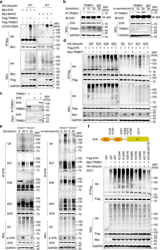
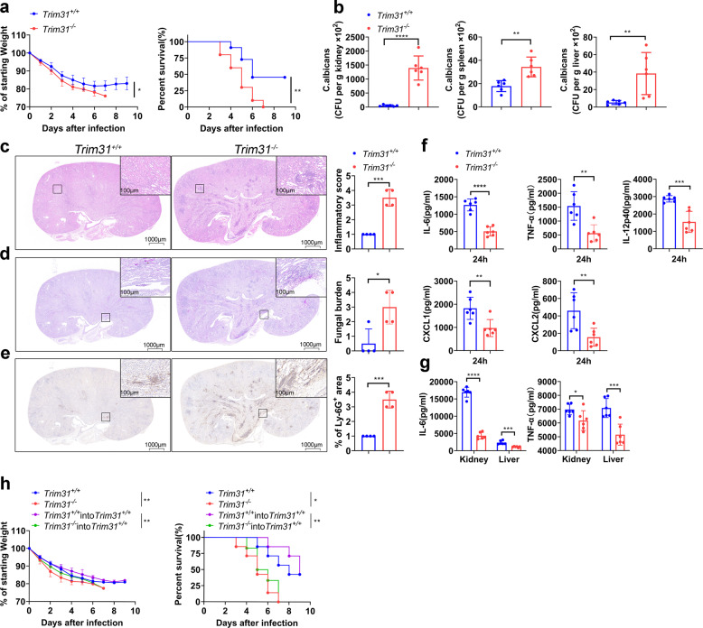
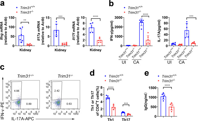
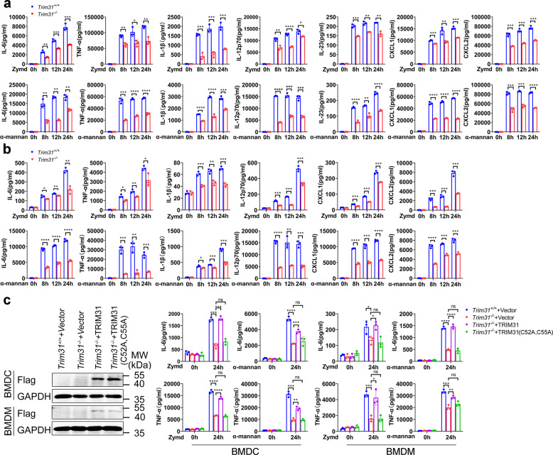
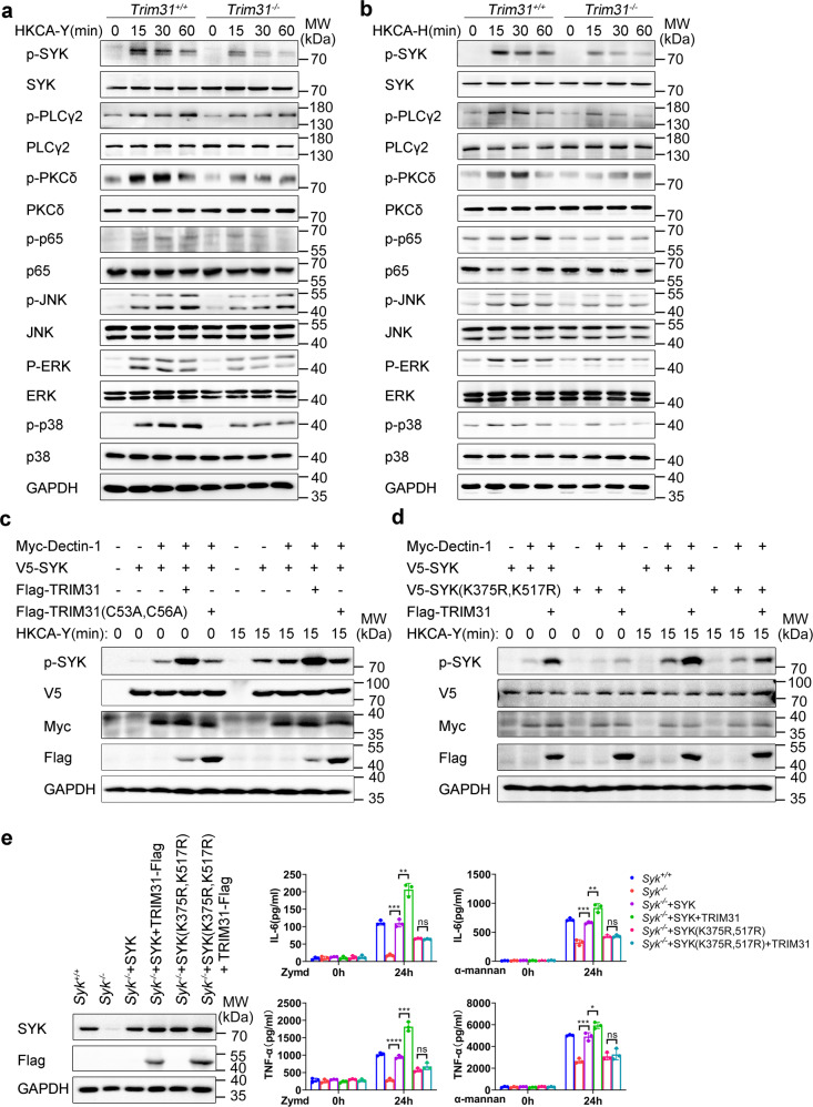
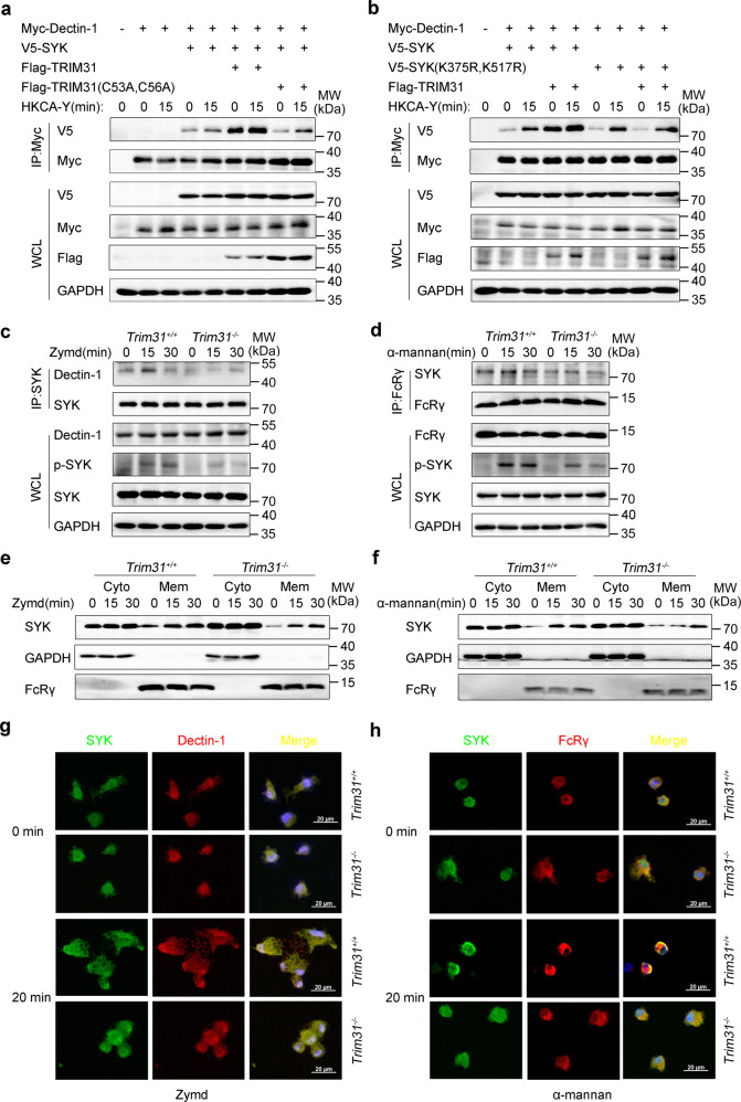
Spleen tyrosine kinase (SYK) is a non-receptor tyrosine kinase, which plays an essential role in both innate and adaptive immunity. However, the key molecular mechanisms that regulate SYK activity are poorly understood. Here we identified the E3 ligase TRIM31 as a crucial regulator of SYK activation. We found that TRIM31 interacted with SYK and catalyzed K27-linked polyubiquitination at Lys375 and Lys517 of SYK. This K27-linked polyubiquitination of SYK promoted its plasma membrane translocation and binding with the C-type lectin receptors (CLRs), and also prevented the interaction with the phosphatase SHP-1. Therefore, deficiency of Trim31 in bone marrow-derived dendritic cells (BMDCs) and macrophages (BMDMs) dampened SYK-mediated signaling and inhibited the secretion of proinflammatory cytokines and chemokines against the fungal pathogen Candida albicans infection. Trim31-/- mice were also more sensitive to C. albicans systemic infection than Trim31+/+ mice and exhibited reduced Th1 and Th17 responses. Overall, our study uncovered the pivotal role of TRIM31-mediated K27-linked polyubiquitination on SYK activation and highlighted the significance of TRIM31 in anti-C. albicans immunity.


Conflict of interest statement

The authors declare no competing interests.

Figures

Fig. 1
Fig. 1
TRIM31 catalyzes the K27-linked polyubiquitination of SYK. a HEK293T cells were transfected with Myc-SYK or Myc-MAVS, HA-ubiquitin (WT), Flag-TRIM31, or Flag-TRIM31 (C53A, C56A) and ubiquitination assays were performed. b Co-IP analysis of the endogenous interaction between TRIM31 and SYK in BMDCs stimulated with Zymd (left) or α-mannan (right) for 0-30 mins. c In vitro Co-IP analysis of TRIM31 with SYK using recombinant TRIM31 and SYK. d ubiquitination assays analysis of SYK in HEK293T cells transfected with Flag-SYK, Myc-TRIM31, and HA-Ubiquitin or its mutants (K6, K11, K27, K29, K33, K48, and K63). e Immunoprecipitation analysis of ubiquitination of endogenous SYK in Trim31+/+ and Trim31−/− BMDCs stimulated with Zymd (left) and α-mannan (right) for indicated time points. f Schematic diagram of lysine residues in SYK for ubiquitination identified by mass spectrometry (top). Immunoprecipitation analysis of ubiquitination of SYK and its mutants in HEK293T cells transfected with Myc-TRIM31, Flag-SYK (WT or mutants) along with HA-Ubiquitin (K27) (bottom). Antibodies (left margins). The above experiments were repeated at least twice with similar results, and the representative data were shown (a–f)
Fig. 2
Fig. 2
TRIM31 is indispensable for anti-fungal immunity. a Weight loss (left) and survival (right) of Trim31+/+ (n = 11), Trim31−/− (n = 10) male littermates (7–8-week-old) after infection with C. albicans strain SC5314 (2 × 105 fungal cells per mouse). b Statistics of C. albicans in the kidneys, spleens, and livers of Trim31+/+ and Trim31−/− mice infected with C. albicans for 5 days, evaluated by serial dilution of homogenized tissues and presented as CFU per gram of the indicated tissue. c Representative images of hematoxylin–eosin (HE)-stained kidney sections of Trim31+/+ and Trim31−/− mice 5 days after systemic C. albicans infection, and obtained inflammatory score based on tissue destruction and renal immune cell infiltration. d Representative images of periodic acid-Schiff (PAS)-stained kidney sections of Trim31+/+ and Trim31−/− mice 5 days after C. albicans infection, and obtained score based on fungal load. e Representative images of kidney sections stained for the neutrophil marker Lys-6G+ and quantification of kidney area scored as Ly-6G+ 5 days after infection of Trim31+/+ and Trim31−/− mice with C. albicans. f ELISA analysis of serum cytokines and chemokines from Trim31+/+ and Trim31−/− mice (n = 6 per group) 24 h after infection with C. albicans (1 × 106 fungal cells per mouse). g ELISA analysis of IL-6 and TNF-α from the supernatant of the organ of the Trim31+/+ and Trim31−/− mice 5 days after systemic C. albicans infection (2 × 105 fungal cells per mouse). h Weight loss (left), as compared to starting weight, and survival (right) over time of lethally irradiated Trim31+/+ mice that were reconstituted with bone marrow from Trim31+/+ (n = 7) or Trim31−/− mice (n = 6), recipient mice were intravenous injected with 1 × 107 BM cells. The control group was Trim31+/+ (n = 7) and Trim31−/− (n = 7). Both of them were subsequently infected with C. albicans (2 × 105 fungal cells per mouse) though the tail vein. *P < 0.05; **P < 0.01; ***P < 0.001 and ****P < 0.0001 (two-way analysis of variance in (a, h left), log-rank test (a, h right) or Student’s t-test (b–g)). One representative experiment of three independent experiments is presented (b–g, Data are shown as mean ± s.d.). Each point represents a single mouse (b, c–g). Representative images of Trim31+/+ (n = 4) and Trim31−/− (n = 4) are shown, three sections per kidney were analyzed, scale bars, 1000 µm, 100 µm (insets) (c–e)
Fig. 3
Fig. 3
TRIM31 is indispensable for anti-fungal Th1 and Th17 responses. a qRT-PCR analysis of Ifng, Il17a, and Il17f from kidneys of Trim31+/+ (n = 6) and Trim31−/− (n = 6) mice infected with C. albicans (2 × 105 fungal cells per mouse) for 5 days. b ELISA analysis of IFN-γ (left) and IL-17A (right) in the supernatant of splenic cells obtained from Trim31+/+ and Trim31−/− mice (n = 6 per group) infected with C. albicans (2 × 105 fungal cells per mouse) for 5 days, followed by stimulation with HKCA-Y (MOI, 1) for 2 days. c, d Splenic cells isolated from the Trim31+/+ or Trim31−/− mice and stimulated as in (b). Intracellular staining of IFN-γ (Th1) and IL-17A (Th17) were determined with flow cytometry. The representative figure is shown in (c), and the results are summarized in (d). e ELISA analysis of IgG in serum from Trim31+/+ and Trim31−/− mice (n = 6 per group) infected with C. albicans (2 × 105 fungal cells per mouse) for 5 days. *P < 0.05; **P < 0.01; ***P < 0.001; and ****P < 0.0001 (Student’s t-test in a, b, d, e). Data are from one experiment representative of three independent experiments (a, b, d, e; mean ± s.d.)
Fig. 4
Fig. 4
TRIM31 controls antifungal activities of BMDCs and BMDMs. a ELISA analysis of IL-6, TNF-α, IL-1β, IL-12, IL-23, CXCL1, and CXCL2 in supernatants of Trim31+/+ and Trim31−/− in BMDCs stimulated with Zymd (100 µg/ml) and α-mannan (100 µg/ml) for the indicated time points. b ELISA analysis of IL-6, TNF-α, IL-1β, IL-12, CXCL1, and CXCL2 in supernatants of Trim31+/+ and Trim31−/− in BMDMs stimulated with Zymd and α-mannan for the indicated time points. The ligands and concentrations are the same as (a). c Immunoblot analysis of the protein expression of TRIM31 in Trim31−/− BMDCs and BMDMs transduced as in right (left). ELISA analysis of IL-6 (upper) and TNF-α (lower) in supernatants of Trim31+/+ and Trim31−/− in BMDCs and BMDMs, respectively, transduced by lentiviral vector GV492, GV492-mTRIM31, and GV492-mTRIM31 (C52A, C55A) followed by stimulation with Zymd and α-mannan for 0–24 h (right). *P < 0.05; **P < 0.01; ***P < 0.001; and ****P < 0.0001 (Student’s t-test in a–c). Experiments were done in triplicates (mean ± s.d. in a–c)
Fig. 5
Fig. 5
TRIM31 promotes SYK phosphorylation. a, b Western blot analysis of phosphorylated and total proteins in lysates of Trim31+/+ and Trim31−/− BMDCs stimulated with HKCA-Y (MOI, 2) (a) or HKCA-H (MOI, 1) (b) for indicated time points. c Immunoblot analysis of SYK phosphorylation and activation in HEK293T cells co-transfected by various combinations of plasmids expressing V5-SYK, Myc-Dectin-1, Flag-TRIM31, and Flag-TRIM31 (C53A, C56A), cells were unstimulated or stimulated by HKCA-Y (MOI, 2) for 15 min. d Immunoblot analysis of SYK or SYK (K375R, K517R) phosphorylation and activation in HEK293T cells transfected by various combinations of plasmids expressing V5-SYK, V5-SYK (K375R, K517R), Myc-Dectin-1, and Flag-TRIM31, cells were unstimulated or stimulated by HKCA-Y (MOI, 2) for 15 min. e Immunoblot analysis of the protein level of SYK or its mutants in Syk−/− BMDCs transduced by the indicated lentiviruses (left). ELISA analysis of IL-6 or TNF-α in Syk−/− BMDCs transduced as in the left panel, followed by stimulation for 0–24 h with Zymd or α-mannan (right). ns, not significant (P > 0.05); *P < 0.05; **P < 0.01; ***P < 0.001; and ****P < 0.0001 (Student’s t-test). Data are representative of three independent experiments (a–e)
Fig. 6
Fig. 6
TRIM31 positively regulates translocation of SYK to the membrane and colocalization with Dectin-1 and FcRγ. a HEK293T cells were co-transfected with Myc-Dectin-1, V5-SYK, Flag-TRIM31, or Flag-TRIM31 (C53A, C56A) by various combinations, cells were unstimulated or stimulated with HKCA-Y (MOI, 2) for 15 min. Then cell lysates were IP with anti-Myc and WCL, respectively, probed with antibodies (left margins). b HEK293T cells were transfected with Myc-Dectin-1, V5-SYK, V5-SYK (K375R, K517R), and Flag-TRIM31 by various combinations, cells were unstimulated or stimulated with HKCA-Y (MOI, 2) for 15 min. Followed by IP with anti-Myc and WCL, respectively, probed with antibodies (left margins). c, d Trim31+/+ and Trim31−/− BMDCs unstimulated (0 min) or stimulated with Zymd (c) or α-mannan (d) for 15 or 30 min, followed by IP with anti-SYK or anti-FcRγ, probed with antibodies (left margins). e, f Trim31+/+ and Trim31−/− BMDCs were stimulated with Zymd (e) or α-mannan (f) for 15 or 30 min, and whole-cell lysates were separated into membrane and cytosolic fractions. Immunoblot analysis with antibodies (left margins). g, h Using SYK-conjugated specific antibody (green), Dectin-1 or FcRγ conjugated specific antibody (red) while the nuclear compartment was observed by DAPI-staining of DNA (blue). Scale bars, 20 µm. Data are from one experiment representative of three independent experiments (a–h)
Fig. 7
Fig. 7
TRIM31 inhibits SHP-1 regulation SYK activation. a, b Trim31+/+ and Trim31−/− BMDCs uninfected (0 min) or infected Zymd (a) or α-mannan (b) for 15 min and 30 min, followed by IP with anti-SYK, probed with anti-SHP-1, WCL immunoblot analysis with indicated antibodies (left margins). c HEK293T cells were transfected with Myc-SYK, V5-SHP-1 and Flag-TRIM31, or Flag-TRIM31 (C53A, C56A). Followed by IP with anti-V5, probed with indicated antibodies (left margins). d HEK293T cells were transfected with Myc-SYK, Myc-Dectin-1, V5-SHP-1, Flag-TRIM31, or Flag-TRIM31 (C53A, C56A) by various combinations, cells were unstimulated or stimulated with HKCA-Y (MOI, 2) for 15 min, WCL immunoblot analysis with indicated antibodies (left margins). e HEK293T cells transfected by various combinations of plasmids expressing Flag-SYK, Flag-SYK (K375R, K517R), Myc-Dectin-1, V5-SHP-1, and Flag-TRIM31, cells were unstimulated or stimulated by HKCA-Y (MOI, 2) for 15 min. WCL immunoblot analysis with indicated antibodies (left margins). Data are from one experiment representative of three independent experiments (a–e)
Fig. 8
Fig. 8
A hypothetical model of the role of TRIM31 in anti-fungal immunity response. TRIM31 catalyzes the K27-linked polyubiquitination of SYK, which can disrupt the autoinhibited status of SYK, promote SYK binding to CLRs, decrease the association of SYK and SHP-1, leading to the increased SYK kinase activity (left). TRIM31 deficiency leads less SYK translocation to the membrane, produces lower amounts of pro-inflammatory cytokines and chemokine in response to C. albicans infection (right)

References

    1. Armstrong-James D, Meintjes G, Brown GD. A neglected epidemic: fungal infections in HIV/AIDS. Trends Microbiol. 2014;22:120–127. doi: 10.1016/j.tim.2014.01.001. - DOI - PubMed
    1. Ravikumar S, Win MS, Chai LY. Optimizing outcomes in immunocompromised hosts: understanding the role of immunotherapy in invasive fungal diseases. Front. Microbiol. 2015;6:1322. - PMC - PubMed
    1. Lanternier F, et al. Primary immunodeficiencies underlying fungal infections. Curr. Opin. Pediatr. 2013;25:736–747. doi: 10.1097/MOP.0000000000000031. - DOI - PMC - PubMed
    1. Zuo T, et al. Alterations in fecal fungal microbiome of patients with COVID-19 during time of hospitalization until discharge. Gastroenterology. 2020;159:1302–1310. doi: 10.1053/j.gastro.2020.06.048. - DOI - PMC - PubMed
    1. Brown GD, et al. Hidden killers: human fungal infections. Sci. Transl. Med. 2012;4:165rv113. doi: 10.1126/scitranslmed.3004404. - DOI - PubMed

Publication types

MeSH terms