. 2021 Aug 6;12(1):4760.
doi: 10.1038/s41467-021-24978-w.

The intervening domain is required for DNA-binding and functional identity of plant MADS transcription factors

Affiliations

The intervening domain is required for DNA-binding and functional identity of plant MADS transcription factors

Xuelei Lai et al. Nat Commun. .

Abstract

The MADS transcription factors (TF) are an ancient eukaryotic protein family. In plants, the family is divided into two main lineages. Here, we demonstrate that DNA binding in both lineages absolutely requires a short amino acid sequence C-terminal to the MADS domain (M domain) called the Intervening domain (I domain) that was previously defined only in type II lineage MADS. Structural elucidation of the MI domains from the floral regulator, SEPALLATA3 (SEP3), shows a conserved fold with the I domain acting to stabilise the M domain. Using the floral organ identity MADS TFs, SEP3, APETALA1 (AP1) and AGAMOUS (AG), domain swapping demonstrate that the I domain alters genome-wide DNA-binding specificity and dimerisation specificity. Introducing AG carrying the I domain of AP1 in the Arabidopsis ap1 mutant resulted in strong complementation and restoration of first and second whorl organs. Taken together, these data demonstrate that the I domain acts as an integral part of the DNA-binding domain and significantly contributes to the functional identity of the MADS TF.


Conflict of interest statement

The authors declare no competing interests.

Figures

Fig. 1
Fig. 1. Structure and sequence of the SEP3 MI domain and corresponding SRF and MEF2 regions.
a Overlay of SEP3 dimer (rainbow) and SRF (1HBX) dimer (grey). The I region (circled in red) is alpha helical for both structures, however the contacts with the M domain differ. N and C-termini are labelled. b Overlay of SEP3 (dark purple) and MEF2A (3KOV, light purple) and SRF (gray) demonstrating the different conformations of the I region in the type II (MEF2-like) and I (SRF-like) MADS TFs, orientation as per a. c SEP3 dimer with one monomer in purple and one in green with N- and C-termini labelled. Amino acids important for interactions between I domains and between M and I domains are labelled and drawn as sticks. Hydrogen bonds are shown as dashed yellow lines. d View of the I domain with intermolecular interactions from the N-terminal alpha helices shown. e Partial sequence alignment of SEP3 (accession NP564214.2), MEF2 (accession AAB25838.1) and SRF (accession P11831) corresponding to the region in the crystal structure of SEP3. Residues directly contacting the DNA for MEF2 and SRF are highlighted in blue.
Fig. 2
Fig. 2. Type I MADS possesses I domain-like region which is required for both dimerisation and DNA binding.
a Amino acid enrichment of the I region (~30 amino acids C-terminal to the M domain) of type I, type II and all MADS TFs, logos generated with WebLogo. The overall height of the stack in each position indicates the sequence information content at that position, while the height of the amino acid symbols within the stack indicates the relative frequency at each position. The MADS TF sequences are taken from The Arabidopsis Information Resource (www.arabidopsis.org). b, c Pull-down assay showing that M domain of AGL61 (AGL61M) does not interact with the M domain of PHE (PHEM) or AGL80 (AGL80M). d, e Pull-down assay showing that the M domain plus the I-like region of AGL61 (AGL61MI) interacts with the MI region of PHE (PHEMI) and AGL80 (AGL80MI). All assays were performed twice and a representative blot is shown. f EMSA assay showing that heterodimers AGL61MI-AGL80MI and AGL61MI-PHEMI shift a DNA sequence containing a canonical CArG-box binding site from the SEP3 promoter, while their corresponding constructs without the I region do not exhibit any binding. All EMSAs were performed at least twice and a representative gel is shown.
Fig. 3
Fig. 3. Type II MADS TFs require the I domain for DNA binding but not dimerisation.
ac Pull-down assay showing that the M domain of SEP3 (SEP3M) interacts with the M domains of AG (AGM), AG with the first 16 N-terminal amino acids deleted (AG(∆N)M) and AP1 (AP1M), respectively. df Pull-down assay showing that the MI domain of SEP3 (SEP3MI) interacts with the MI domain of AGMI, AG(∆N)MI and AP1MI, respectively. All assays were performed twice and a representative blot is shown. g, h EMSA assay showing that homodimers from SEP3MI, AGMI, AG(ΔN)MI and AP1MI and heterodimers SEP3MI-AGMI, SEP3MI-AG(∆N)MI and SEP3MI-AP1MI shift a DNA sequence containing a canonical CArG-box binding site (as per Fig. 2), while their corresponding constructs without the I domain cannot, suggesting that the I domain in type II MADS TFs is required for DNA binding. All EMSAs were performed at least twice and a representative gel is shown.
Fig. 4
Fig. 4. DNA-binding and protein interactions patterns.
a Comparison of SEP3-AG and SEP3-AGIAP1 seq-DAP-seq binding intensity (log10 of reads per kb per million of reads mapped in bound regions) and colour coded by purple-blue (SEP3-AGIAP1 specific) to orange-red (SEP3-AG specific) according to log10 of SEP3-AGIAP1/SEP3-AG. b Density plot showing data as per a. c Logos derived from PWM-based models obtained for SEP3-AG and SEP3-AGIAP1. d Predictive power of TFBS models. Models are built using 600 sequences best bound by each of the two heterocomplexes and are searched against 1073 SEP3-AG (orange) and 1073 SEP3-AGIAP1 (blue) specific regions, defined as the top 15% of sequences that are most strongly bound by one complex relative to the other. Matrix-based models (PWM and TFFM) are not able to differentiate SEP3-AG and SEP3-AGIAP1 binding, whereas k-mer-based analysis is able to better predict binding for the respective datasets. e SEP3-AG favours intersite spacings of 47 and 57 bp based on SEP3-AG-specific regions. SEP3-AGIAP1 favours intersite spacings of 25 and 34 bp based on SEP3-AGIAP1I specific regions. f Top, published SELEX-seq for SEP3-AP1 and SEP3-AG comparing the normalised score ratios (SEP3-AG/SEP3-AP1) for SELEX-seq and score ratios (AG/AP1) ChIP-seq at 1500 SEP3 best bound loci in ChIP-seq show a positive correlation, suggesting that SEP3-AP1 and SEP-AG bind different sequences in vivo and that in vitro binding is able to differentiate bound sequences that are more SEP3-AP1-like versus SEP3-AG-like. Bottom, SEP3-AG and SEP3-AGIAP1 seq-DAP-seq coverage as per SELEX-seq scores. A positive correlation is observed suggesting that, in vitro, the swap of AP1 I domain in AG is able to recover some of the binding specificity of SEP3-AP1. g Yeast two-hybrid assays using AG, AP1 and AGIAP1 as bait against MIKCC MADS TFs in Arabidopsis. Data show that AGIAP1 loses AG interactors and gains AP1 interactors.
Fig. 5
Fig. 5. Primary transformants in the ap1-11 mutant background exhibit a spectrum of complementation.
a Schematic of the constructs and proteins produced with AP1 protein in purple and AG in dark blue. Domains are labelled MIKC. b Flower phenotypes of T1 transformants (n ≥ 10). All transformants were in the ap1-11 background. Phenotypes for WT and ap1-11 are shown. The domains corresponding to AP1 and AG are coloured as per a and shown schematically below each panel labelled 1–6 in the top left. The number of primary transformants exhibiting a given phenotype is listed in the top right of panel 1–6.
Fig. 6
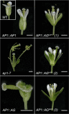
Fig. 6. AP1:AGIAP1 expression largely complements the ap1-7 flower phenotype.
ap1-7 lines expressing either AP1 (AP1::AP1), AG (AP1::AG) or AGIPA1 (AP1::AGIAP1) under the control of the AP1 promoter were grown at 22 °C in long days. A typical WT flower is shown as an insert in the upper left panel. Representative flowers for three independent AP1::AGIAP1 lines are presented and one representative flower from AP1::AG and AP1::AP1 expressing lines. While the first whorl in ap1-7 is replaced by bract or stipule with axillary buds and petals are missing in the second whorl, AP1::AGIAP1 expression restores WT first and second whorl organs, while AG expression triggers carpel development in the first whorl and absence of petals in the second whorl. Scales bars = 1 mm.
Fig. 7
Fig. 7. First and second whorls cell identity are complemented in ap1-7 plants expressing AGIAP1.
First (a) and second (b) whorl organs were removed from WT, and flowers from AP1 or AGIAP1 expressing ap1-7 plants. First whorl organs in AGIAP1 expressing plants are slightly longer compared to WT and AP1 expressing plants (a, left panel). Second whorl organs in AGIAP1 expressing plants are slightly smaller compared to WT and AP1 expressing plants (b, left panel). Epidermal cell identity, observed by SEM, shows characteristic WT elongated sepal cells in AP1 and AGIAP1 expressing plants (a, right panel) and characteristic WT conical cells in petals from AP1 and AGIAP1 expressing plants (b, right panel). A small number of epidermal cells typical of leaves are also seen in AGIAP1 expressing plants and to a lesser extent in AP1 expressing plants (arrow). Scale bars indicate 100 µM for SEM images and 1 mm for organ photographs.

References

    1. Passmore S, Maine GT, Elble R, Christ C, Tye BK. Saccharomyces cerevisiae protein involved in plasmid maintenance is necessary for mating of MAT alpha cells. J. Mol. Biol. 1988;204:593–606. doi: 10.1016/0022-2836(88)90358-0. - DOI - PubMed
    1. Yanofsky MF, et al. The protein encoded by the Arabidopsis homeotic gene agamous resembles transcription factors. Nature. 1990;346:35–39. doi: 10.1038/346035a0. - DOI - PubMed
    1. Sommer H, et al. Deficiens, a homeotic gene involved in the control of flower morphogenesis in Antirrhinum majus: the protein shows homology to transcription factors. EMBO J. 1990;9:605–613. doi: 10.1002/j.1460-2075.1990.tb08152.x. - DOI - PMC - PubMed
    1. Norman C, Runswick M, Pollock R, Treisman R. Isolation and properties of cDNA clones encoding SRF, a transcription factor that binds to the c-fos serum response element. Cell. 1988;55:989–1003. doi: 10.1016/0092-8674(88)90244-9. - DOI - PubMed
    1. Martinez-Castilla LP, Alvarez-Buylla ER. Adaptive evolution in the Arabidopsis MADS-box gene family inferred from its complete resolved phylogeny. Proc. Natl Acad. Sci. USA. 2003;100:13407–13412. doi: 10.1073/pnas.1835864100. - DOI - PMC - PubMed

Publication types

MeSH terms