. 2021 Aug 6;11(1):16036.
doi: 10.1038/s41598-021-95396-7.

Equine pituitary pars intermedia dysfunction: a spontaneous model of synucleinopathy

Affiliations

Equine pituitary pars intermedia dysfunction: a spontaneous model of synucleinopathy

Jessica S Fortin et al. Sci Rep. .

Abstract

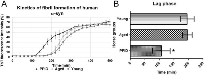
Equine pituitary pars intermedia dysfunction (PPID) is a common endocrine disease of aged horses that shows a similar pathophysiology as Parkinson's Disease (PD) with increased levels of α-synuclein (α-syn). While α-syn is thought to play a pathogenic role in horses with PPID, it is unclear if α-syn is also misfolded in the pars intermedia and could similarly promote self-aggregation and propagation. Consequently, α-syn was isolated from the pars intermedia from groups of healthy young and aged horses, and aged PPID-afflicted horses. Seeding experiments confirmed the prion-like properties of α-syn isolated from PPID-afflicted horses. Next, detection of α-syn fibrils in pars intermedia via transmission electron microscopy (TEM) was exclusive to PPID-afflicted horses. A bank of fragment peptides was designed to further characterize equine α-syn misfolding. Region 62-87 of equine and human α-syn peptides was found to be most prone to aggregation according to Tango bioinformatic program and kinetics of aggregation via a thioflavin T fluorescence assay. In both species, fragment peptide 62-87 is capable of generating mature fibrils as demonstrated by TEM. The combined animal, bioinformatic, and biophysical studies provide evidence that equine α-syn is misfolded in PPID horses.


Conflict of interest statement

The authors declare no competing interests.

Figures

Figure 1
Figure 1
Alpha-synuclein (α-syn) extracted from the pars intermedia of PPID-affected horses has the potential to cross seed recombinant human α-syn. (A) Thioflavin T (ThT) fluorescence assay kinetics of human α-syn fibril formation in the presence of equine α-syn isolated from young, aged, and PPID-affected horses. Data represent the mean of the fluorescence intensity with error bars for SEM obtained from three different horses in each group (young, aged, PPID). (B) Lag phase time (min) for initiation of human α-syn fibril formation during incubation with equine α-syn extracted from pars intermedia tissue collected from young, aged and PPID-affected horses. The time (min) required for human α-syn fibril elongation (lag phase) is shorter when induced with equine α-syn extracted from PPID-affected horses. Data represent the mean of the lag phase time (min) obtained from five different horses in each group (young, aged, PPID). Data were analyzed by the one-way analysis of variance with Dunnett's multiple comparison post-hoc testing between groups. (*significant difference p < 0.05.)
Figure 2
Figure 2
Alpha-synuclein (α-syn) extracted from the pars intermedia of PPID-affected horses exhibits a fibrillar ultrastructure as visualized by transmission electron microscopy (TEM). (A) TEM image of the background staining showing the absence of fibrils in equine α-syn isolated by immunoprecipitation (IP) from the pars intermedia of a young horse. (Scale bar, 100 nm.) (B) TEM image of α-syn isolated by IP from pars intermedia of an aged horse representing background staining (no fibrils). (Scale bar, 100 nm.) (C) TEM image of positively stained equine α-syn fibrils isolated by IP from the pars intermedia of a PPID-affected horse. (Scale bar, 100 nm.) (D) TEM image of a collagen fiber. (Scale bar, 200 nm.)
Figure 3
Figure 3
Immunogold transmission electron microscopy (TEM) using an α-synuclein primary antibody on pars intermedia tissue confirms presence of fibrils in a PPID-affected horse. (A) TEM image from the pars intermedia of an PPID-affected horse without use of the primary antibody consists of background staining of the secondary antibody. (B) TEM image of the pars intermedia of an aged horse shows an immunogold labeling similar to background straining. (C) TEM image of the pars intermedia of a PPID-affected horse exhibits immunogold labeling. (D) TEM image of the pars intermedia of a PPID-affected horse contains a fibril with minimal immunogold labeling in the extracellular milieu (Scale bars, 200 nm).
Figure 4
Figure 4
Equine α-synuclein (α-syn) fragment peptide 62–86 generated fibrils after a longer lag phase period in comparison with its human counterpart using ThT binding assays. (A) Sequence of α-syn synthetic fragment peptides used to investigate aggregation propensity of equine α-syn. Aggregation scores determined by Tango bioinformatic program are provided in the right-sided column for each peptide fragment. Fragments exhibiting an aggregation score of 30–60 were selected as negative controls. Fragments with scores near or above 300 were further analyzed with biophysical assays to study the propensity of aggregation in vitro. (B) The maximum of fluorescence intensity at plateau phase of selected fragment peptides: bars show mean of fluorescence with error bars for SEM of three replicates. (C) Kinetics of α-syn fibril formation of human and equine fragment peptides 62–86. Data represent the mean of the fluorescence intensity with error bars for SEM obtained from three replicates. (D) Lag time required for fibril elongation of equine fragment peptide 62–86 is longer than for the human fragment peptide 62–86. Two-way analysis of variance with Bonferonni post-hoc testing to compare replicate means by row. Differences were considered statistically significant at * p < 0.05 and ** p < 0.01.
Figure 5
Figure 5
Transmission electron microscopic (TEM) images of human and equine α-synuclein (α-syn) peptide fragments at the end of kinetics of fibril formation (after 96 h of incubation at 37 °C). (A) TEM images of human α-syn full-length recombinant peptide at the end of the kinetics of aggregation. (Scale bar, 200 nm.) (B) TEM image of human and equine α-syn synthetic fragment 1–25. (Scale bar, 100 nm.) (C) TEM image of human and equine α-syn synthetic fragment 26–50. (Scale bar, 100 nm.) (D) TEM image of human α-syn synthetic fragment 37–61. (Scale bar, 200 nm.) (E) TEM image of equine α-syn synthetic fragment 37–61. (Scale bar, 200 nm.) (F) TEM image of human α-syn synthetic fragment 62–86. (Scale bar, 200 nm.) (G) TEM image of equine α-syn synthetic fragment 62–86. (Scale bar, 200 nm.)

References

    1. Marras C, et al. Prevalence of Parkinson's disease across North America. N.P.J. Parkinsons. Dis. 2018;4:21. doi: 10.1038/s41531-018-0058-0. - DOI - PMC - PubMed
    1. Casajus Pelegay E, Puzzo F, Yilmazer A, Cagin U. Targeting mitochondrial defects to increase longevity in animal models of neurodegenerative diseases. Adv. Exp. Med. Biol. 2019;1134:89–110. doi: 10.1007/978-3-030-12668-1_5. - DOI - PubMed
    1. Dawson TM, Golde TE, Lagier-Tourenne C. Animal models of neurodegenerative diseases. Nat. Neurosci. 2018;21(10):1370–1379. doi: 10.1038/s41593-018-0236-8. - DOI - PMC - PubMed
    1. Fernandez CI, Lopez J, Martinez L. Non-clinical models for neurodegenerative diseases: therapeutic approach and drug validation in animal models. Behav. Sci. (Basel) 2017;7(4):82. doi: 10.3390/bs7040082. - DOI - PMC - PubMed
    1. Guyer C. Recent developments in animal models of human neurodegenerative diseases. Toxicol. Pathol. 2000;28(2):363–366. doi: 10.1177/019262330002800218. - DOI - PubMed

Publication types

Substances