Review
. 2022 Jan;47(1):3-19.
doi: 10.1038/s41386-021-01076-5. Epub 2021 Aug 6.

Evolution of prefrontal cortex

Affiliations
Review

Evolution of prefrontal cortex

Todd M Preuss et al. Neuropsychopharmacology. 2022 Jan.

Abstract

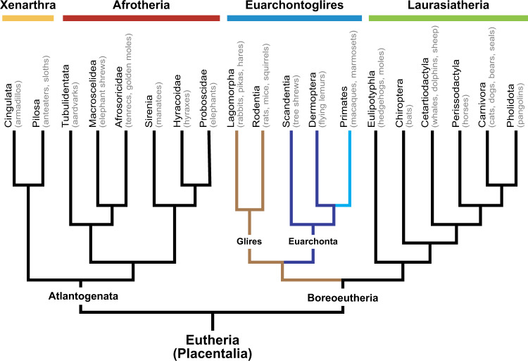
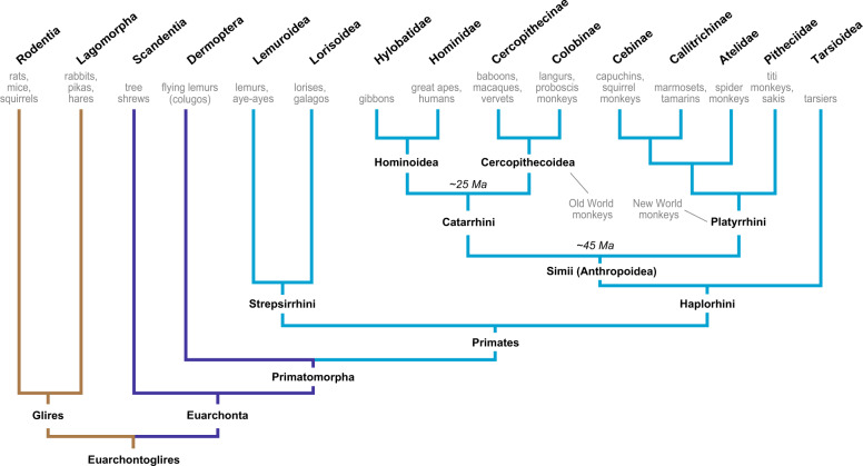
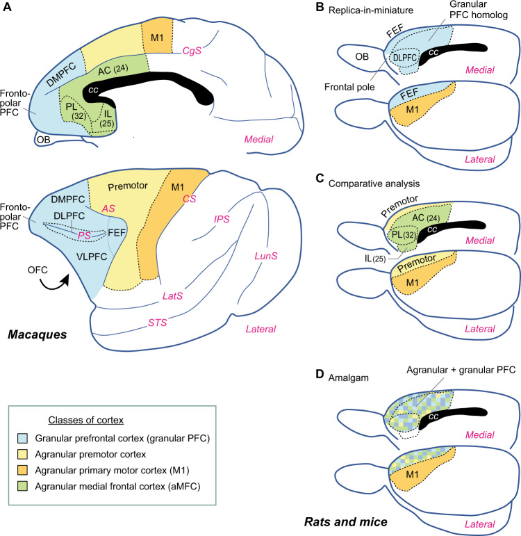
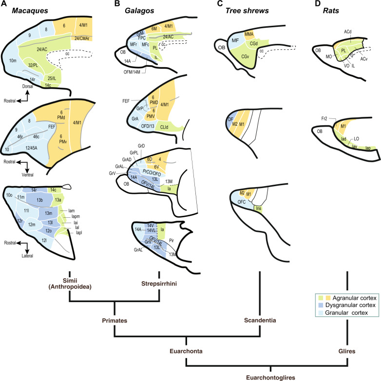
Subdivisions of the prefrontal cortex (PFC) evolved at different times. Agranular parts of the PFC emerged in early mammals, and rodents, primates, and other modern mammals share them by inheritance. These are limbic areas and include the agranular orbital cortex and agranular medial frontal cortex (areas 24, 32, and 25). Rodent research provides valuable insights into the structure, functions, and development of these shared areas, but it contributes less to parts of the PFC that are specific to primates, namely, the granular, isocortical PFC that dominates the frontal lobe in humans. The first granular PFC areas evolved either in early primates or in the last common ancestor of primates and tree shrews. Additional granular PFC areas emerged in the primate stem lineage, as represented by modern strepsirrhines. Other granular PFC areas evolved in simians, the group that includes apes, humans, and monkeys. In general, PFC accreted new areas along a roughly posterior to anterior trajectory during primate evolution. A major expansion of the granular PFC occurred in humans in concert with other association areas, with modifications of corticocortical connectivity and gene expression, although current evidence does not support the addition of a large number of new, human-specific PFC areas.


Conflict of interest statement

The authors declare no competing interests.

Figures

Fig. 1
Fig. 1. A phylogenetic tree of placental mammals, based on molecular phylogenomics.
The best-established supraordinal clades are labeled at the top. Adapted from Murphy et al. [12, 327].
Fig. 2
Fig. 2. Relationships among Euarchontoglires [12, 28].
Estimated times for the catarrhine–platyrrhine and the hominoid–cercopithecoid divergences appear in italics. Ma million years ago.
Fig. 3
Fig. 3. Proposed homologies among frontal areas in primates and rodents.
Areas shaded with the same color have been advanced as homologs. A Macaque brains serve as representative simians for comparison with rodents. The curved arrow indicates the location of OFC on the hidden, ventral surface of the macaque frontal lobe. B An example of the idea that rodents have some or all of the granular PFC areas observed in simians, albeit in miniature form. C The view advocated here, which is based on comparative neuroanatomy. D The amalgam theory, in which alternating colored voxels indicate the intermixing of areas. AC anterior cingulate cortex (area 24 in primates), aMFC agranular medial frontal cortex, AS arcuate sulcus, cc corpus callosum, CgS cingulate sulcus, CS central sulcus, DLPFC dorsolateral PFC (also known as the periprincipal PFC), DMPFC dorsomedial PFC, FEF frontal eye field, IL infralimbic cortex (area 25 in primates), IPS intraparietal sulcus, LatS lateral sulcus, LunS lunate sulcus, M1 primary motor cortex, OB olfactory bulb, PFC prefrontal cortex, OFC orbitofrontal cortex, VLPFC ventrolateral PFC, PL prelimbic cortex (area 32 in primates), PS principal sulcus, STS superior temporal sulcus. Adapted from Preuss and Robert [6].
Fig. 4
Fig. 4. Types of cortical areas in the frontal cortex of Euarchontoglires.
A Macaques (Macaca) as representative simians. Adapted from Carmichael and Price [328]. B Galagos (Otolemur) as representative strepsirrhines. This is an amalgamation of the interpretations of Preuss and Goldman-Rakic [203] and Wong and Kaas [205]. C Tree shrews (Tupaia) as representative non-primate Euarchontans. Adapted from Wong and Kaas [198]. D Rats (Rattus) as representative Glires. Adapted from Palomero-Gallagher and Zilles [329]. Labeling has been retained from the originals to the extent possible. a or A anterior or a subdivision of an area (versus b or B), AC anterior cingulate cortex, c caudal, cc corpus callosum, CG cingulate-gyrus cortex, CLI claustral isocortex, CMA cingulate motor area, d or D dorsal, DF dorsal frontal area, FPC frontopolar cortex, Fr2 second frontal area, also known as the medial agranular area, Gr granular, i inferior, Ia agranular insular cortex, IL infralimbic cortex, Ins insular cortex, l or L lateral, LO lateral orbitofrontal cortex, m or M medial, M1 primary motor cortex, M2, in this case, another part of M1 or a premotor area (not homologous with M2 in simians or rodents, which, in turn, are not homologous with each other), MF medial frontal area, MMA medial motor area, MO medial orbitofrontal cortex, o or O orbital, OB olfactory bulb, OF orbitofrontal cortex, OFO opercular orbitofrontal cortex, p posterior, Pir piriform cortex, PL prelimbic cortex, PM premotor cortex, PrCO opercular proisocortex, r rostral, v or V ventral, VO ventral orbitofrontal cortex.
Fig. 5
Fig. 5. Enlargement of the granular PFC in human evolution.
A Granular PFC size relative to the remainder of the frontal lobe. Solid line: regression; dashed lines: confidence limits. B Phylogenetic statistical tests reveal that the increase in hominids (blue) is significant. C Percentage of granular PFC within the frontal lobe. D Phylogenetic statistics show that the clade including chimpanzees and humans underwent a significantly greater increase in relative granular PFC volume contrasted with other homotypical association areas, namely those in the parietal and temporal lobes. A, B, D Reproduced, with permission (RightsLink license 5035380571023, license date March 24, 2021) from Smaers et al. [282]. C Plotted from data in Elston et al. [330]. PFC prefrontal cortex.

Similar articles

Cited by

References

    1. Manger PR, Cort J, Ebrahim N, Goodman A, Henning J, Karolia M, et al. Is 21st century neuroscience too focussed on the rat/mouse model of brain function and dysfunction? Front Neuroanat. 2008;2:5. - PMC - PubMed
    1. Bolker J. Model organisms: there’s more to life than rats and flies. Nature. 2012;491:31–3. - PubMed
    1. Logan CA. The altered rationale for the choice of a standard animal in experimental psychology: Henry H. Donaldson, Adolf Meyer and ‘the’ albino rat. Hist Psychol. 1999;2:3–24. - PubMed
    1. Murray EA, Wise SP, Graham KS. The evolution of memory systems: ancestors, anatomy, and adaptations. Oxford, UK: Oxford University Press; 2017.
    1. Preuss TM. The discovery of cerebral diversity: an unwelcome scientific revolution. In: Falk D, Gibson KR, editors. Evolutionary anatomy of the primate cerebral cortex. Cambridge, UK: Cambridge University Press; 2001. p. 138–64.

Publication types