

3D T1-weighted turbo spin echo contrast-enhanced MRI at 1.5 T for frameless brain metastases radiotherapy
- PMID: 34363123
- PMCID: PMC11800802
- DOI: 10.1007/s00432-021-03755-8
3D T1-weighted turbo spin echo contrast-enhanced MRI at 1.5 T for frameless brain metastases radiotherapy
Abstract
Purpose: Performance of 3D-T1W-TSE has been proven superior to 3D-MP-GRE at 3 T on brain metastases (BM) contrast-enhanced (CE) MRI. However, its performance at 1.5 T is largely unknown and sparsely reported. This study aims to assess image quality, lesion detectability and conspicuity of 1.5 T 3D-T1W-TSE on planning MRI of frameless BM radiotherapy.
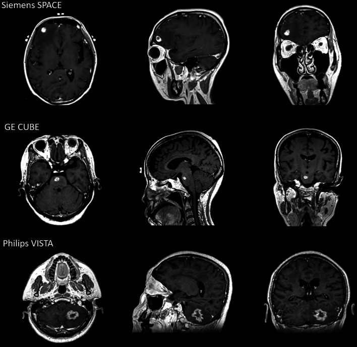
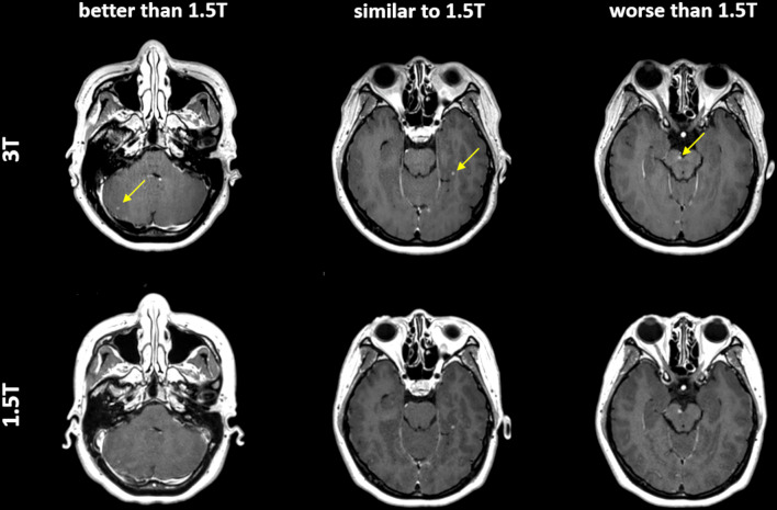
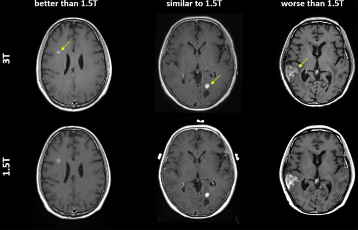
Methods: 94 BM patients to be treated by frameless brain radiotherapy were scanned using 3D-T1W-TSE with immobilization on multi-vendor 1.5 T MRI-simulators. BMs were jointly diagnosed by 4 reviewers. Enhanced lesion conspicuity was quantitatively assessed by calculating contrast ratio (CR) and contrast-to-noise ratio (CNR). Signal-to-noise ratio (SNR) reduction of white matter due to the use of flexible coil was assessed. Lesion detectability and conspicuity were compared between 1.5 T planning MRI and 3 T diagnostic MRI by an oncologist and a radiologist in 10 patients.
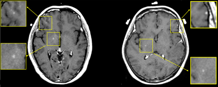
Results: 497 BMs were jointly diagnosed. The CR and CNR were 75.2 ± 39.9% and 14.2 ± 8.1, respectively. SNR reduced considerably from 31.7 ± 8.3 to 21.9 ± 5.4 with the longer distance to coils. 3 T diagnostic MRI and 1.5 T planning MRI yielded exactly the same detection of 84 BMs. Qualitatively, lesion conspicuity at 1.5 T was not inferior to that at 3 T. Quantitatively, lower brain SNR and lesion CNR were found at 1.5 T, while lesion CR at 1.5 T was highly comparable to that at 3 T.
Conclusion: 1.5 T 3D-T1W-TSE planning MRI of frameless BM radiotherapy was comprehensively assessed. Highly comparable BM detectability and conspicuity were achieved by 1.5 T planning MRI compared to 3 T diagnostic MRI. 1.5 T 3D-T1W-TSE should be valuable for frameless brain radiotherapy planning.
Keywords: 3D T1-weighted turbo spin echo; Brain metastases; Contrast enhanced MRI; MR-guided-radiotherapy; Stereotactic radiotherapy.
© 2021. The Author(s), under exclusive licence to Springer-Verlag GmbH Germany, part of Springer Nature.
Conflict of interest statement
The authors have no relevant conflicts of interest to disclose.
Figures
References
-
- Bednarz G, Downes MB, Corn BW, Curran WJ, Goldman HW (1999) Evaluation of the spatial accuracy of magnetic resonance imaging-based stereotactic target localization for gamma knife radiosurgery of functional disorders. Neurosurgery 45:1156–1161. 10.1097/00006123-199911000-00028 - PubMed
MeSH terms
Substances
Grants and funding
LinkOut - more resources
Full Text Sources
Medical
