

Three-dimensional microengineered vascularised endometrium-on-a-chip
- PMID: 34363466
- PMCID: PMC8450871
- DOI: 10.1093/humrep/deab186
Three-dimensional microengineered vascularised endometrium-on-a-chip
Abstract
Study question: Can we reconstitute physiologically relevant 3-dimensional (3D) microengineered endometrium in-vitro model?
Summary answer: Our representative microengineered vascularised endometrium on-a-chip closely recapitulates the endometrial microenvironment that consists of three distinct layers including epithelial cells, stromal fibroblasts and endothelial cells in a 3D extracellular matrix in a spatiotemporal manner.
What is known already: Organ-on-a-chip, a multi-channel 3D microfluidic cell culture system, is widely used to investigate physiologically relevant responses of organ systems.
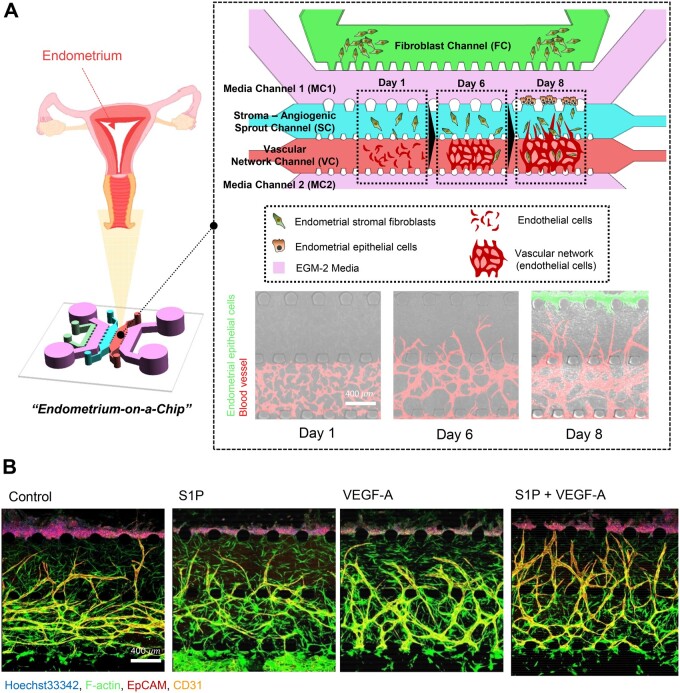
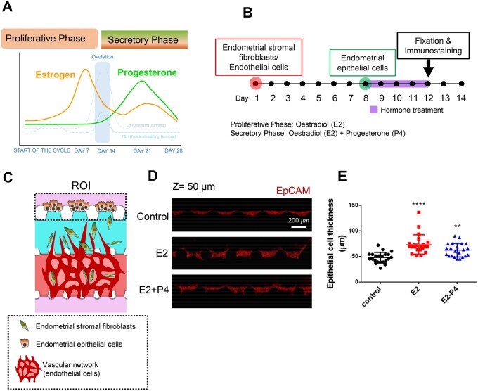
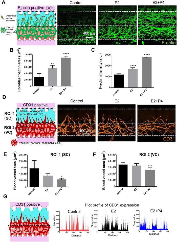
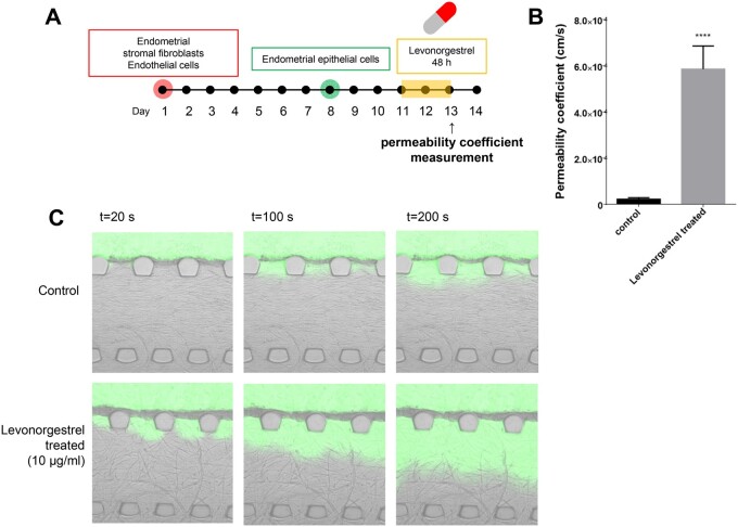
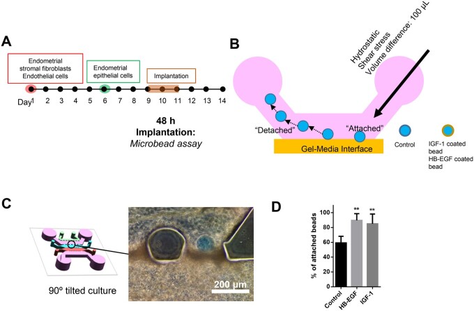
Study design, size, duration: The device consists of five microchannels that are arrayed in parallel and partitioned by array of micropost. Two central channels are for 3D culture and morphogenesis of stromal fibroblast and endothelial cells. In addition, the outermost channel is for the culture of additional endometrial stromal fibroblasts that secrete biochemical cues to induce directional pro-angiogenic responses of endothelial cells. To seed endometrial epithelial cells, on Day 8, Ishikawa cells were introduced to one of the two medium channels to adhere on the gel surface. After that, the microengineered endometrium was cultured for an additional 5-6 days (total ∼ 14 days) for the purpose of each experiment.
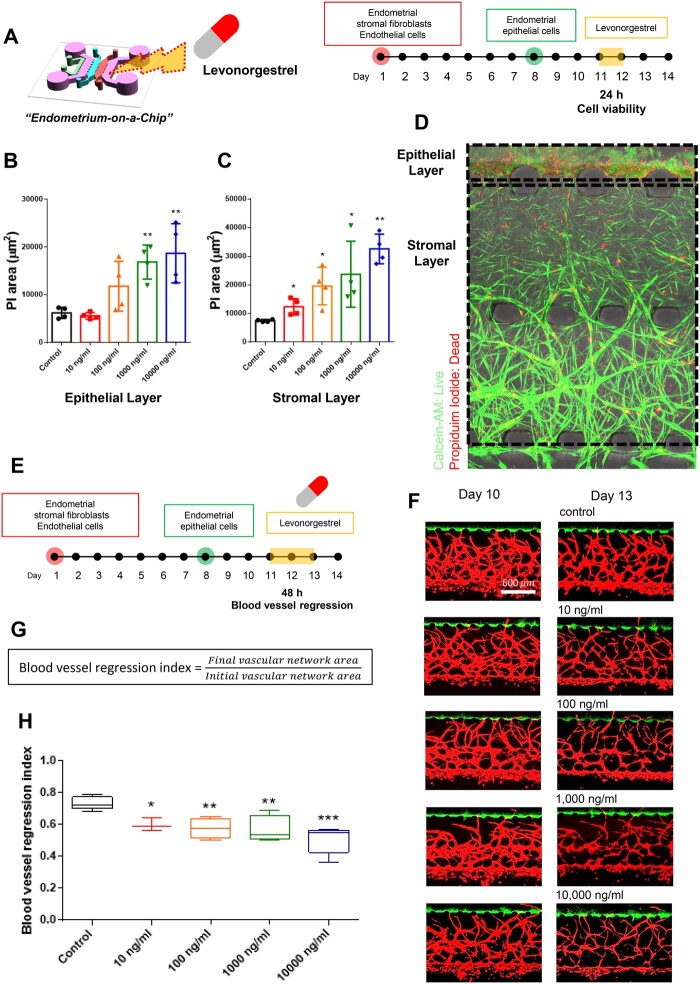
Participants/materials, setting, methods: Microfluidic 3D cultures were maintained in endothelial growth Medium 2 with or without oestradiol and progesterone. Some cultures additionally received exogenous pro-angiogenic factors. For the three distinct layers of microengineered endometrium-on-a-chip, the epithelium, stroma and blood vessel characteristics and drug response of each distinct layer in the microfluidic model were assessed morphologically and biochemically. The quantitative measurement of endometrial drug delivery was evaluated by the permeability coefficients.
Main results and the role of chance: We established microengineered vascularised endometrium-on-chip, which consists of three distinct layers: epithelium, stroma and blood vessels. Our endometrium model faithfully recapitulates in-vivo endometrial vasculo-angiogenesis and hormonal responses displaying key features of the proliferative and secretory phases of the menstrual cycle. Furthermore, the effect of the emergency contraception drug levonorgestrel was evaluated in our model demonstrating increased endometrial permeability and blood vessel regression in a dose-dependent manner. We finally provided a proof of concept of the multi-layered endometrium model for embryo implantation, which aids a better understanding of the molecular and cellular mechanisms underlying this process.
Large scale data: N/A.
Limitations, reasons for caution: This report is largely an in-vitro study and it would be beneficial to validate our findings using human primary endometrial cells.
Wider implications of the findings: Our 3D microengineered vascularised endometrium-on-a-chip provides a new in-vitro approach to drug screening and drug discovery by mimicking the complicated behaviours of human endometrium. Thus, we suggest our model as a tool for addressing critical challenges and unsolved problems in female diseases, such as endometriosis, uterine cancer and female infertility, in a personalised manner.
Study funding/competing interest(s): This work is supported by funding from the National Research Foundation of Korea (NRF) grant funded by the Korea government (MSIT) to Y.J.K. (No. 2018R1C1B6003), to J.A. (No. 2020R1I1A1A01074136) and to H.S.K. (No. 2020R1C1C100787212). The authors report no conflicts of interest.
Keywords: 3D culture; drug screening; endometrial angiogenesis; endometrium; microfluidic.
© The Author(s) 2021. Published by Oxford University Press on behalf of European Society of Human Reproduction and Embryology.
Figures
References
-
- Ahn J, Cho CS, Cho SW, Kang JH, Kim SY, Min DH, Song JM, Park TE, Jeon NL.. Investigation on vascular cytotoxicity and extravascular transport of cationic polymer nanoparticles using perfusable 3D microvessel model. Acta Biomater 2018;76:154–163. - PubMed
-
- Ahn J, Ko J, Lee S, Yu J, Kim Y, Jeon NL.. Microfluidics in nanoparticle drug delivery; from synthesis to pre-clinical screening. Adv Drug Deliv Rev 2018;128:29–53. - PubMed
-
- Aplin JD.Molecular and cellular aspects of peri-implantation processes, Boston, Massachusetts 15–18 July 1994. Placenta 1995;16:109–111. - PubMed
-
- Brar AK, Frank GR, Kessler CA, Cedars MI, Handwerger S.. Progesterone-dependent decidualization of the human endometrium is mediated by cAMP. Endocrine 1997;6:301–307. - PubMed
Publication types
MeSH terms
LinkOut - more resources
Full Text Sources
Other Literature Sources
Medical
