Systematic review and meta-analysis of chest radiograph (CXR) findings in COVID-19
- PMID: 34364071
- PMCID: PMC8313779
- DOI: 10.1016/j.clinimag.2021.06.039
Systematic review and meta-analysis of chest radiograph (CXR) findings in COVID-19
Abstract
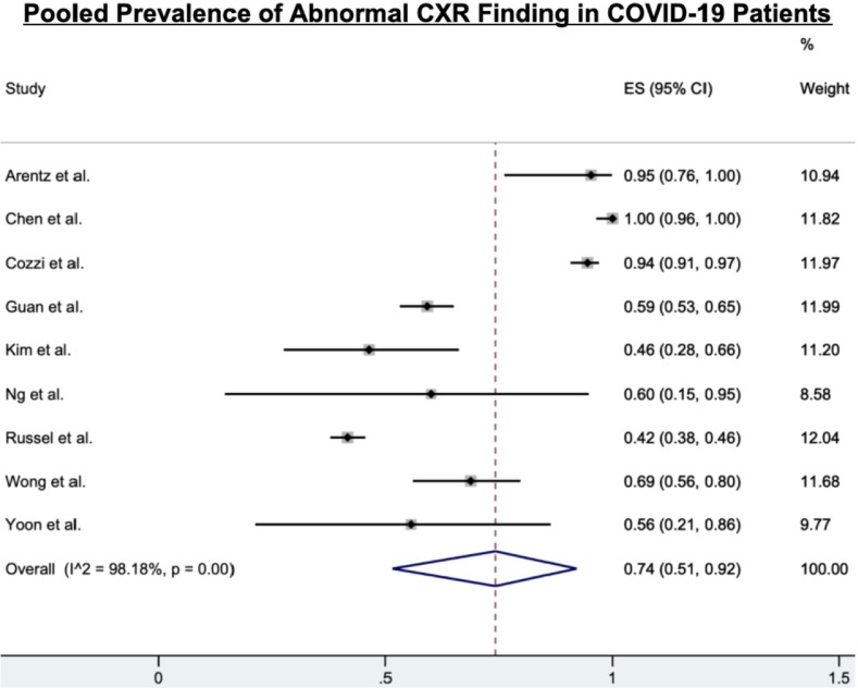
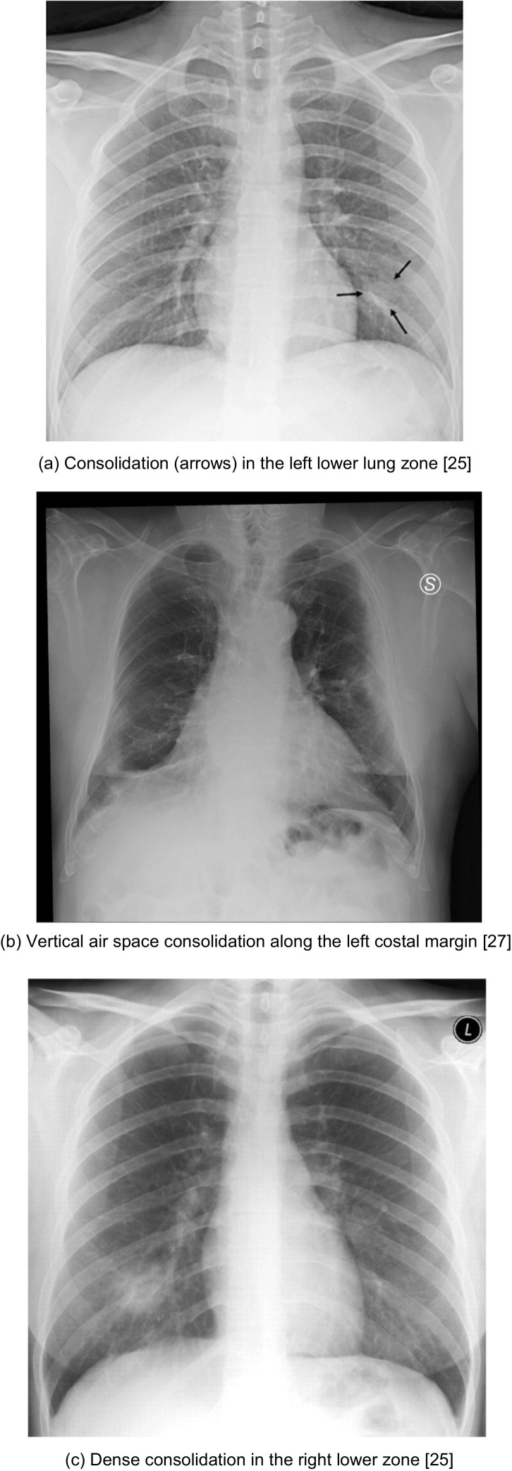
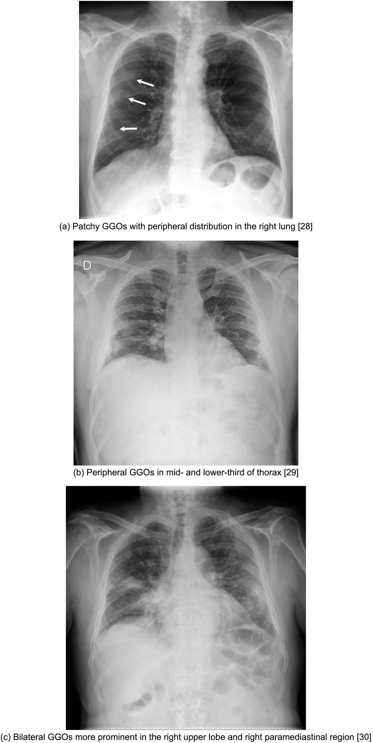
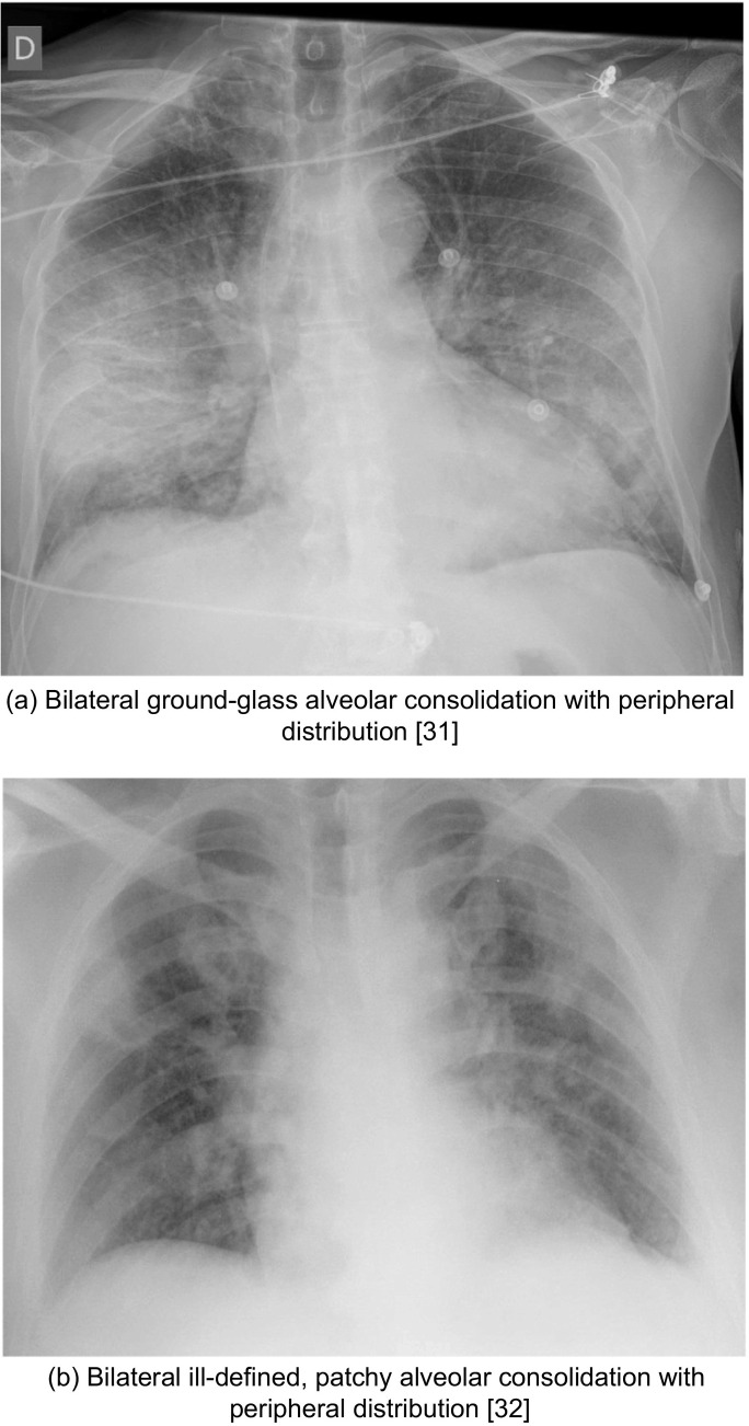
Chest radiography (CXR) is most likely to be the utilized modality for diagnosing COVID-19 and following up on any lung-associated abnormalities. This review provides a meta-analysis of the current literature on CXR imaging findings to determine the most common appearances of lung abnormalities in COVID-19 patients in order to equip medical researchers and healthcare professionals in their efforts to combat this pandemic. Twelve studies met the inclusion criteria and were analyzed. The inclusion criteria consisted of: (1) published in English literature; (2) original research study; (3) sample size of at least 5 patients; (4) reporting clinical characteristics of COVID-19 patients as well as CXR imaging features; and (5) noting the number of patients with each corresponding imaging feature. A total of 1948 patients were included in this study. To perform the meta-analysis, a random-effects model calculated the pooled prevalence and 95% confidence intervals of abnormal CXR imaging findings. Seventy-four percent (74%) (95% CI: 51-92%) of patients with COVID-19 had an abnormal CXR at the initial time of diagnosis or sometime during the disease course. While there was no single feature on CXR that was diagnostic of COVID-19 viral pneumonia, a characteristic set of findings were obvious. The most common abnormalities were consolidation (28%, 95% CI: 8-54%) and ground-glass opacities (29%, 95% CI: 10-53%). The distribution was most frequently bilateral (43%, 95% CI: 27-60%), peripheral (51%, 95% CI: 36-66%), and basal zone (56%, 95% CI: 37-74%) predominant. Contrary to parenchymal abnormalities, pneumothorax (1%, 95% CI: 0-3%) and pleural effusions (6%, 95% CI: 1-16%) were rare.
Keywords: COVID-19; CXR; Chest radiograph; Coronavirus; Imaging; Meta-analysis; SARS-CoV-2.
Copyright © 2021 The Authors. Published by Elsevier Inc. All rights reserved.
Figures


Similar articles
-
Signs and symptoms to determine if a patient presenting in primary care or hospital outpatient settings has COVID-19.Cochrane Database Syst Rev. 2022 May 20;5(5):CD013665. doi: 10.1002/14651858.CD013665.pub3. Cochrane Database Syst Rev. 2022. PMID: 35593186 Free PMC article.
-
Symptom- and chest-radiography screening for active pulmonary tuberculosis in HIV-negative adults and adults with unknown HIV status.Cochrane Database Syst Rev. 2022 Mar 23;3(3):CD010890. doi: 10.1002/14651858.CD010890.pub2. Cochrane Database Syst Rev. 2022. PMID: 35320584 Free PMC article.
-
Antibody tests for identification of current and past infection with SARS-CoV-2.Cochrane Database Syst Rev. 2022 Nov 17;11(11):CD013652. doi: 10.1002/14651858.CD013652.pub2. Cochrane Database Syst Rev. 2022. PMID: 36394900 Free PMC article.
-
Measures implemented in the school setting to contain the COVID-19 pandemic.Cochrane Database Syst Rev. 2022 Jan 17;1(1):CD015029. doi: 10.1002/14651858.CD015029. Cochrane Database Syst Rev. 2022. Update in: Cochrane Database Syst Rev. 2024 May 2;5:CD015029. doi: 10.1002/14651858.CD015029.pub2. PMID: 35037252 Free PMC article. Updated.
-
Thoracic imaging tests for the diagnosis of COVID-19.Cochrane Database Syst Rev. 2022 May 16;5(5):CD013639. doi: 10.1002/14651858.CD013639.pub5. Cochrane Database Syst Rev. 2022. PMID: 35575286 Free PMC article.
Cited by
-
Gastrointestinal symptoms among COVID-19 patients presenting to a primary health care center of Nepal: A cross-sectional study.Health Sci Rep. 2023 Sep 14;6(9):e1568. doi: 10.1002/hsr2.1568. eCollection 2023 Sep. Health Sci Rep. 2023. PMID: 37720170 Free PMC article.
-
Automated prediction of COVID-19 severity upon admission by chest X-ray images and clinical metadata aiming at accuracy and explainability.Sci Rep. 2023 Mar 14;13(1):4226. doi: 10.1038/s41598-023-30505-2. Sci Rep. 2023. PMID: 36918593 Free PMC article.
-
Current and Emerging Knowledge in COVID-19.Radiology. 2023 Feb;306(2):e222462. doi: 10.1148/radiol.222462. Epub 2023 Jan 10. Radiology. 2023. PMID: 36625747 Free PMC article. Review.
-
Molecular imaging as a tool for evaluation of COVID-19 sequelae - A review of literature.World J Radiol. 2022 Jul 28;14(7):194-208. doi: 10.4329/wjr.v14.i7.194. World J Radiol. 2022. PMID: 36160629 Free PMC article. Review.
-
Multireader evaluation of radiologist performance for COVID-19 detection on emergency department chest radiographs.Clin Imaging. 2022 Feb;82:77-82. doi: 10.1016/j.clinimag.2021.10.018. Epub 2021 Nov 12. Clin Imaging. 2022. PMID: 34798562 Free PMC article.
References
-
- Coronavirus (COVID-19) events as they happen. https://www.who.int/emergencies/diseases/novel-coronavirus-2019/events-a...
-
- ACR Recommendations for the use of Chest Radiography and Computed Tomography (CT) for Suspected COVID-19 Infection. https://www.acr.org/Advocacy-and-Economics/ACR-Position-Statements/Recom...
Publication types
MeSH terms
LinkOut - more resources
Full Text Sources
Medical
Miscellaneous