

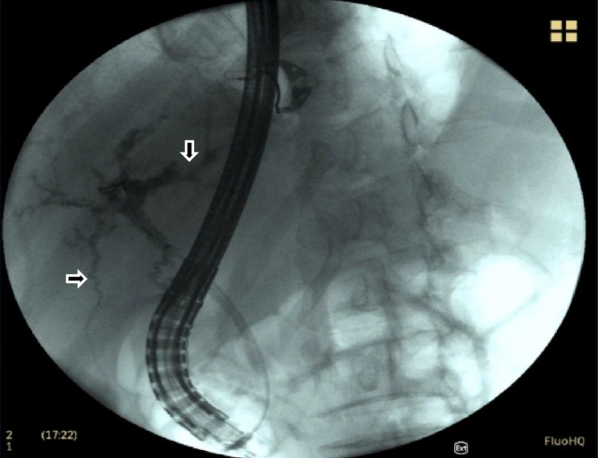
A case of secondary sclerosing cholangitis due to COVID-19
- PMID: 34364072
- PMCID: PMC8314797
- DOI: 10.1016/j.clinimag.2021.07.017
A case of secondary sclerosing cholangitis due to COVID-19
Abstract
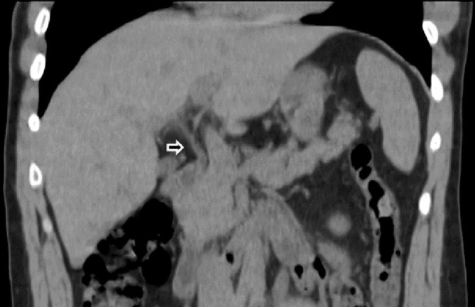
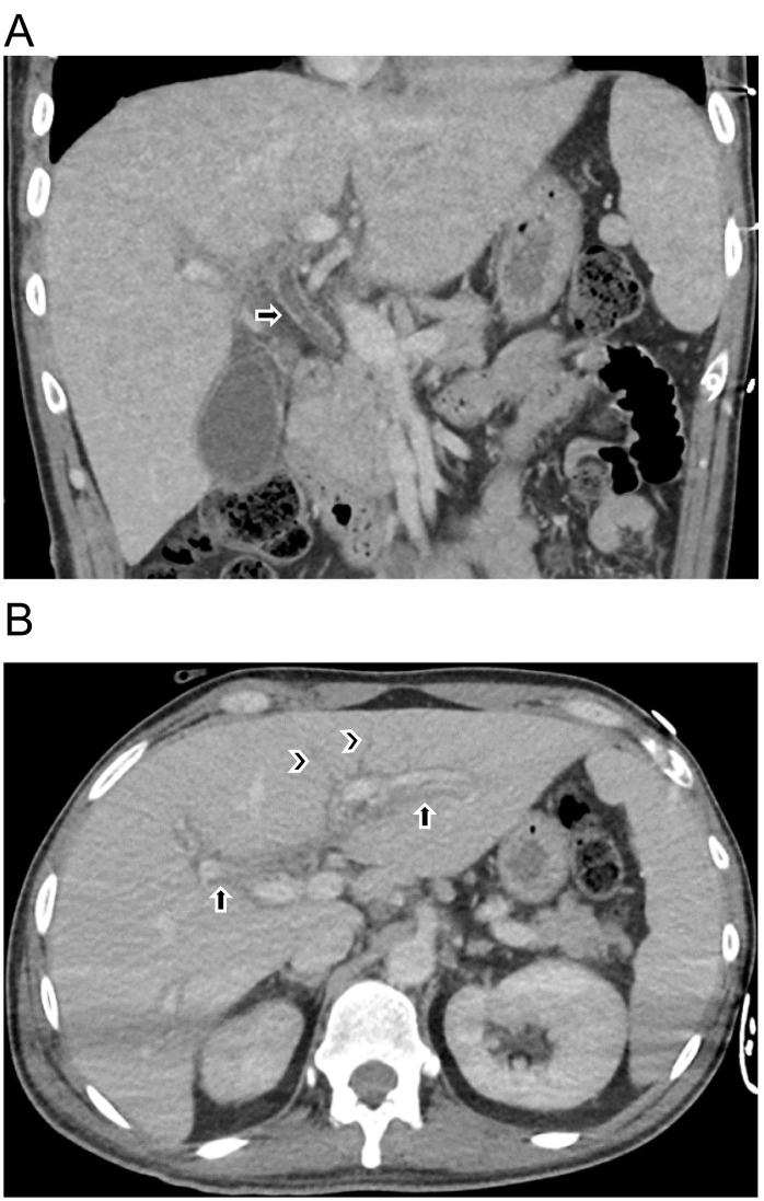
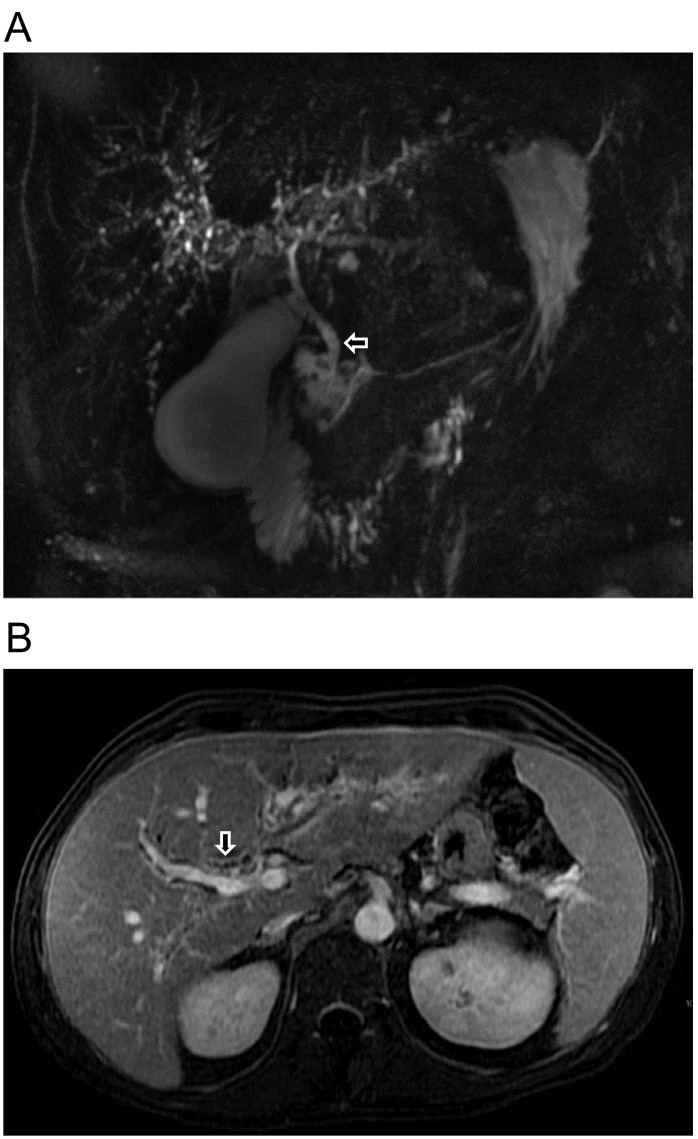
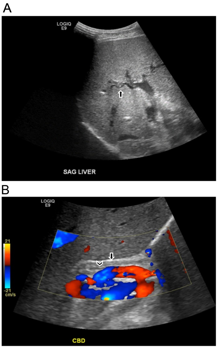
COVID-19 was first recognized by the World Health Organization (WHO) in December 2019 and declared a global pandemic in March 2020. Although COVID-19 primarily results in pulmonary symptoms, it is becoming apparent that it can lead to multisystemic manifestations. Liver damage with elevated AST and ALT is seen in patients with COVID-19. Although the etiology of liver damage is still debated, biliary damage is rarely seen. This case demonstrates a potential complication of COVID-19 in a previously healthy patient. The patient contracted COVID-19 in March 2020 and endured a complicated course including intubation, multiple readmissions, and chronic abdominal pain. He is now awaiting a liver transplant. Our case portrays biliary damage as an additional possible complication of COVID-19 and the importance of imaging in its diagnosis.
Keywords: COVID-19; Cholangiopathy; Hepatobiliary; Secondary sclerosing cholangitis.
Copyright © 2021 Elsevier Inc. All rights reserved.
Figures
References
-
- CDC . June 10, 2021. United States COVID-19 Cases, Deaths, and Laboratory Testing (NAATs) by State, Territory, and Jurisdiction. https://covid.cdc.gov/covid-data-tracker/#cases_casesper100klast7days.https://covid.cdc.gov/covid-data-tracker/#cases_casesper100klast7days Published.
Publication types
MeSH terms
LinkOut - more resources
Full Text Sources
Medical
