A Review of Mechano-Biochemical Models for Testing Composite Restorations
- PMID: 34365857
- PMCID: PMC8716139
- DOI: 10.1177/00220345211026918
A Review of Mechano-Biochemical Models for Testing Composite Restorations
Abstract
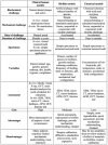
Due to the severe mechano-biochemical conditions in the oral cavity, many dental restorations will degrade and eventually fail. For teeth restored with resin composite, the major modes of failure are secondary caries and fracture of the tooth or restoration. While clinical studies can answer some of the more practical questions, such as the rate of failure, fundamental understanding on the failure mechanism can be obtained from laboratory studies using simplified models more effectively. Reviewed in this article are the 4 main types of models used to study the degradation of resin-composite restorations, namely, animal, human in vivo or in situ, in vitro biofilm, and in vitro chemical models. The characteristics, advantages, and disadvantages of these models are discussed and compared. The tooth-restoration interface is widely considered the weakest link in a resin composite restoration. To account for the different types of degradation that can occur (i.e., demineralization, resin hydrolysis, and collagen degradation), enzymes such as esterase and collagenase found in the oral environment are used, in addition to acids, to form biochemical models to test resin-composite restorations in conjunction with mechanical loading. Furthermore, laboratory tests are usually performed in an accelerated manner to save time. It is argued that, for an accelerated multicomponent model to be representative and predictive in terms of both the mode and the speed of degradation, the individual components must be synchronized in their rates of action and be calibrated with clinical data. The process of calibrating the in vitro models against clinical data is briefly described. To achieve representative and predictive in vitro models, more comparative studies of in vivo and in vitro models are required to calibrate the laboratory studies.
Keywords: biofilms; calibration; composite resins; dental restoration failures; in vitro testing; models.
Conflict of interest statement
Figures


References
-
- Armstrong S, Breschi L, Özcan M, Pfefferkorn F, Ferrari M, Van Meerbeek B. 2017. Academy of Dental Materials guidance on in vitro testing of dental composite bonding effectiveness to dentin/enamel using micro-tensile bond strength (μTBS) approach. Dent Mater. 33(2):133–143. - PubMed
-
- Armstrong SR, Jessop JL, Vargas MA, Zou Y, Qian F, Campbell JA, Pashley DH. 2006. Effects of exogenous collagenase and cholesterol esterase on the durability of the resin-dentin bond. J Adhe Dent. 8(3):151–160. - PubMed
-
- Askar H, Krois J, Gostemeyer G, Bottenberg P, Zero D, Banerjee A, Schwendicke F. 2020. Secondary caries: what is it, and how it can be controlled, detected, and managed? Clin Oral Invest. 24(5):1869–1876. - PubMed
-
- Bista B, Sadr A, Nazari A, Shimada Y, Sumi Y, Tagami J. 2013. Nondestructive assessment of current one-step self-etch dental adhesives using optical coherence tomography. J Biomed Opt. 18(7):76020. - PubMed
-
- Bowen WH. 2013. Rodent model in caries research. Odontology. 101(1):9–14. - PubMed
Publication types
MeSH terms
Substances
Grants and funding
LinkOut - more resources
Full Text Sources
Medical

