Review
. 2021;83(3):939-961.
doi: 10.3233/JAD-210268.

NLRP3 Inflammasome: A Starring Role in Amyloid-β- and Tau-Driven Pathological Events in Alzheimer's Disease

Affiliations
Review

NLRP3 Inflammasome: A Starring Role in Amyloid-β- and Tau-Driven Pathological Events in Alzheimer's Disease

Mariana Van Zeller et al. J Alzheimers Dis. 2021.

Abstract

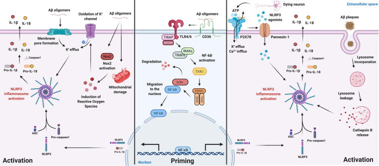
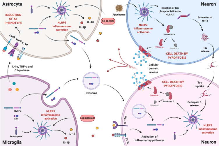
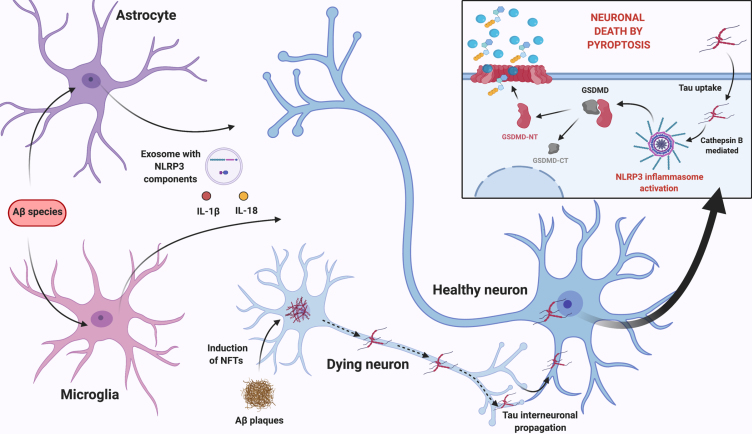
Alzheimer's disease (AD) is the most prevalent neurodegenerative disease commonly diagnosed among the elderly population. AD is characterized by the loss of synaptic connections, neuronal death, and progressive cognitive impairment, attributed to the extracellular accumulation of senile plaques, composed by insoluble aggregates of amyloid-β (Aβ) peptides, and to the intraneuronal formation of neurofibrillary tangles shaped by hyperphosphorylated filaments of the microtubule-associated protein tau. However, evidence showed that chronic inflammatory responses, with long-lasting exacerbated release of proinflammatory cytokines by reactive glial cells, contribute to the pathophysiology of the disease. NLRP3 inflammasome (NLRP3), a cytosolic multiprotein complex sensor of a wide range of stimuli, was implicated in multiple neurological diseases, including AD. Herein, we review the most recent findings regarding the involvement of NLRP3 in the pathogenesis of AD. We address the mechanisms of NLRP3 priming and activation in glial cells by Aβ species and the potential role of neurofibrillary tangles and extracellular vesicles in disease progression. Neuronal death by NLRP3-mediated pyroptosis, driven by the interneuronal tau propagation, is also discussed. We present considerable evidence to claim that NLRP3 inhibition, is undoubtfully a potential therapeutic strategy for AD.

Keywords: Alzheimer’s disease; MCC950; NLRP3; amyloid-β; astrocytes; inflammasome; microglia; neuroinflammation; pro-inflammatory cytokines; pyroptosis; tau.


Conflict of interest statement

Authors’ disclosures available online (https://www.j-alz.com/manuscript-disclosures/21-0268r1).

Figures

Fig. 1
Fig. 1
Schematic illustration for Aβ-mediated NLRP3 inflammasome priming and activation mechanisms described in microglia. Aβ species can work either as a priming stimulus (middle panel) or as an activating stimulus (left and right panels). As a priming signal, Aβ oligomers bind to the CD36 surface receptor, triggering the formation of a TLR4-TLR6 heterodimer. This heterodimer activates a cascade of signaling molecules resulting in the activation and nucleus translocation of the transcription factor NF-kB, that promotes the transcription of NLRP3 domain and of pro-IL-1β. Aβ plaques can act as an activating signal by two main mechanisms (right panel). Aβ is known to cause synaptic dysfunction and neuronal damage. Considering this, P2X7R is activated by ATP released from dying neurons and recruits the Pannexin-1 channel that allows the entrance of NLRP3 agonists to the cell. Also, ATP binding to the purinergic receptor induces K+ efflux and Ca2 + influx, known to promote NLRP3 activation. On the other hand, Aβ plaques can also be phagocytized and incorporated into lysosomes, boosting lysosomal destabilization and consequent content release. Cathepsin B, a lysosomal proteolytic enzyme, promotes the assembly of the inflammasome by a still unknown mechanism. Aβ oligomers are also able to activate NLRP3 through a mechanism that does not involve phagocytosis (left panel). Soluble oligomeric Aβ species can induce pore formation in the cell membrane and ROS production, which then promotes oxidation of K+ channels. These events might culminate in K+ efflux promoting the activation of the inflammasome. The assembly and activation of NLRP3, independently of the mechanism involved, ultimately results in the production and release of the inflammatory cytokines IL-1β and IL-18. Created with BioRender.com.
Fig. 2
Fig. 2
The intercellular communication between neurons and glial cells in the presence of Aβ species is a complex and reciprocal process. Aβ-induced reactive microglia release IL-1α, TNF-α, and C1q, which can be received by resting astrocytes promoting the reactive A1 state. NLRP3 activation in reactive glial cells, boosts the release of pro-inflammatory cytokines IL-1β and IL-18, which can bind to their respective receptors in neurons activating inflammatory pathways. Reactive glial cells might also release NLRP3 components, namely NLRP3 domain and ASC, through exosomes that can be internalized by neurons. On the other hand, Aβ plaques can, in neighboring neurons, mediate the occurrence of pyroptosis and promote the development of NFTs, via NLRP3 activation. More importantly, tau can be released and taken up by other neurons, activating NLRP3 in the recipient cell, hypothetically through lysosomal destabilization and Cathepsin B release, and inducing pyroptosis. Therefore, proximity to Aβ plaques will not determine neuronal death by NLRP3-mediated pyroptosis, since, due to tau interneuronal propagation, pyroptosis can occur in neurons far from plaques. Ultimately, release of cellular content caused by pyroptosis will feed a self-propagating loop of neuroinflammatory mediators. Created with BioRender.com.
Fig. 3
Fig. 3
Proposed multicellular feedback loop that feeds on NLRP3 inflammasome activation in glial cells and neurons. Aβ-induced NLRP3 activation in glial cells, prompts the release of pro-inflammatory cytokines, as well as exosomal release of NLRP3 domain and ASC. This neuroinflammatory milieu sums up to the neurotoxic effect of Aβ plaques in nearby neurons, which induce tau destabilization and NFTs formation, via NLRP3 activation. According to tau interneuronal propagation hypothesis, tau is then released and taken up by neighboring healthy neurons activating NLRP3 in these cells, most likely through lysosomal release of Cathepsin B, causing neuronal death by pyroptosis with cellular content release and perpetuating glial cells’ activation. Created with BioRender.com.

Similar articles

Cited by

References

    1. Bali J, Gheinani AH, Zurbriggen S, Rajendran L (2012) Role of genes linked to sporadic Alzheimer’s disease risk in the production of β-amyloid peptides. Proc Natl Acad Sci U S A 109, 15307–15311. - PMC - PubMed
    1. Masters CL, Bateman R, Blennow K, Rowe CC, Sperling RA, Cummings JL (2015) Alzheimer’s disease. Nat Rev Dis Prim 1, 15056. - PubMed
    1. Stelzmann RA, Norman Schnitzlein H, Reed Murtagh F (1995) An English translation of Alzheimer’s 1907 paper, “über eine eigenartige erkankung der hirnrinde.”. Clin Anat 8, 429–431. - PubMed
    1. Querfurth HW, Laferla FM (2010) Alzheimer’s disease. N Engl J Med 9, 329–344. - PubMed
    1. Hardy J, Allsop D (1991) Amyloid deposition as the central event in the aetiology of Alzheimer’s disease. Trends Pharmacol Sci 12, 383–388. - PubMed

Publication types

Substances