

Presence of Vessel Wall Hyperintensity in Unruptured Arteriovenous Malformations on Vessel Wall Magnetic Resonance Imaging: Pilot Study of AVM Vessel Wall "Enhancement"
- PMID: 34366779
- PMCID: PMC8334001
- DOI: 10.3389/fnins.2021.697432
Presence of Vessel Wall Hyperintensity in Unruptured Arteriovenous Malformations on Vessel Wall Magnetic Resonance Imaging: Pilot Study of AVM Vessel Wall "Enhancement"
Abstract
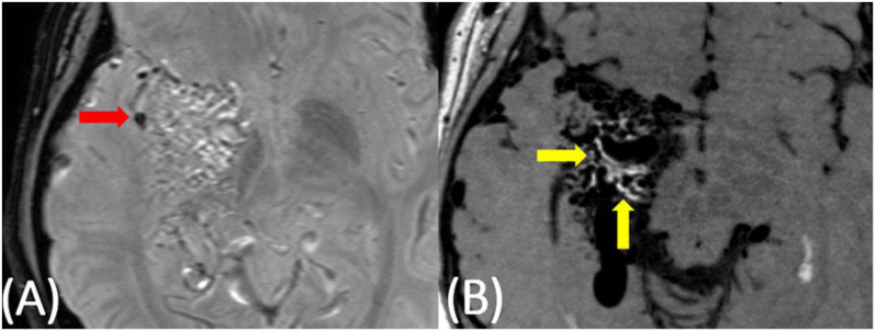
Purpose: High-resolution vessel wall magnetic resonance imaging (VW-MRI) could provide a way to identify high risk arteriovenous malformation (AVM) features. We present the first pilot study of clinically unruptured AVMs evaluated by high-resolution VW-MRI. Methods: A retrospective review of clinically unruptured AVMs with VW-MRI between January 1, 2016 and December 31, 2018 was performed documenting the presence or absence of vessel wall "hyperintensity," or enhancement, within the nidus as well as perivascular enhancement and evidence of old hemorrhage (EOOH). The extent of nidal vessel wall "hyperintensity" was approximated into five groups: 0, 1-25, 26-50, 51-75, and 76-100%. Results: Of the nine cases, eight demonstrated at least some degree of vessel wall nidus "hyperintensity." Of those eight cases, four demonstrated greater than 50% of the nidus with hyperintensity at the vessel wall, and three cases had perivascular enhancement adjacent to nidal vessels. Although none of the subjects had prior clinical hemorrhage/AVM rupture, of the six patients with available susceptibility weighted imaging to assess for remote hemorrhage, only two had subtle siderosis to suggest prior sub-clinical bleeds. Conclusion: Vessel wall "enhancement" occurs in AVMs with no prior clinical rupture. Additional studies are needed to further investigate the implication of these findings.
Keywords: MRI; arteriovenous malformation; unruptured AVM; vessel wall enhancement; vessel wall imaging.
Copyright © 2021 Eisenmenger, Junn, Cooke, Hetts, Zhu, Johnson, Manunga, Saloner, Hess and Kim.
Conflict of interest statement
The authors declare that the research was conducted in the absence of any commercial or financial relationships that could be construed as a potential conflict of interest.
Figures


References
-
- Boussel L., Rayz V., McCulloch C., Martin A., Acevedo-Bolton G., Lawton M., et al. (2008). Aneurysm growth occurs at region of low wall shear stress: patient-specific correlation of hemodynamics and growth in a longitudinal study. Stroke 39 2997–3002. 10.1161/strokeaha.108.521617 - DOI - PMC - PubMed
-
- Dieleman N., van der Kolk A. G., Zwanenburg J. J., Harteveld A. A., Biessels G. J., Luijten P. R., et al. (2014). Imaging intracranial vessel wall pathology with magnetic resonance imaging: current prospects and future directions. Circulation 130 192–201. 10.1161/circulationaha.113.006919 - DOI - PubMed