

Low-Frequency TMS Results in Condition-Related Dynamic Activation Changes of Stimulated and Contralateral Inferior Parietal Lobule
- PMID: 34366812
- PMCID: PMC8342925
- DOI: 10.3389/fnhum.2021.684367
Low-Frequency TMS Results in Condition-Related Dynamic Activation Changes of Stimulated and Contralateral Inferior Parietal Lobule
Abstract
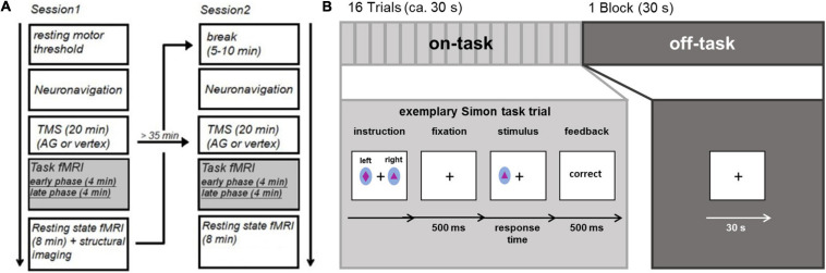
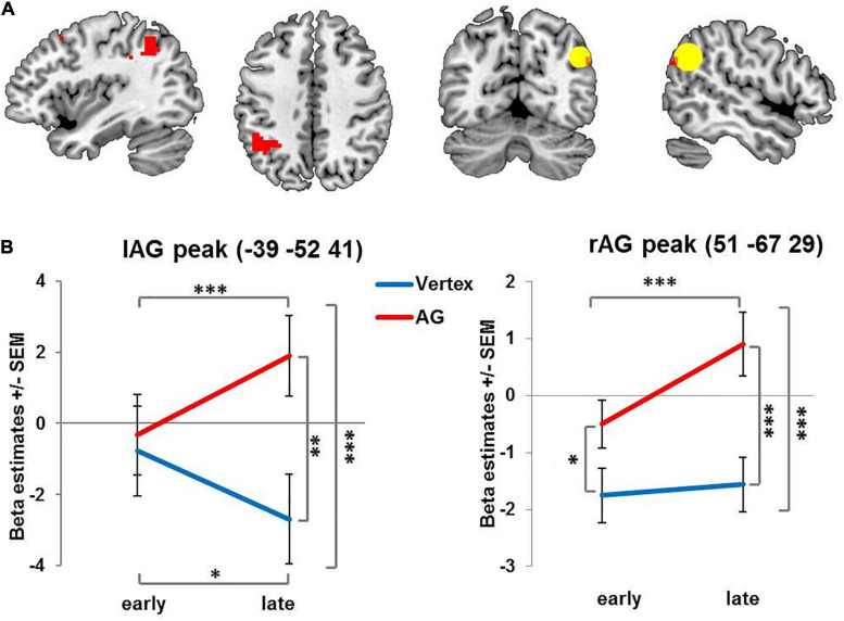
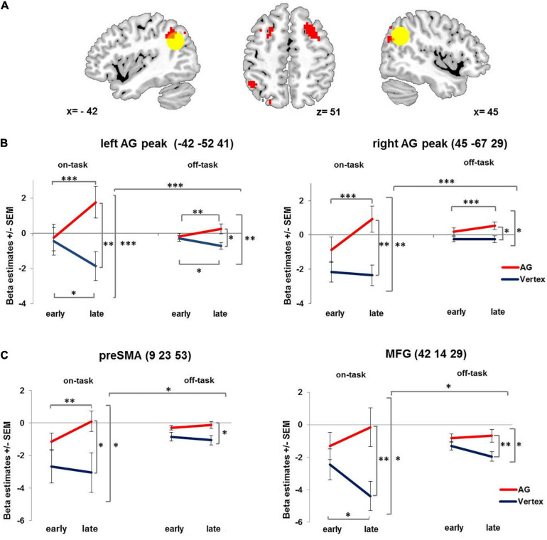
Non-invasive brain stimulation is a promising approach to study the causal relationship between brain function and behavior. However, it is difficult to interpret behavioral null results as dynamic brain network changes have the potential to prevent stimulation from affecting behavior, ultimately compensating for the stimulation. The present study investigated local and remote changes in brain activity via functional magnetic resonance imaging (fMRI) after offline disruption of the inferior parietal lobule (IPL) or the vertex in human participants via 1 Hz repetitive transcranial magnetic stimulation (rTMS). Since the IPL acts as a multimodal hub of several networks, we implemented two experimental conditions in order to robustly engage task-positive networks, such as the fronto-parietal control network (on-task condition) and the default mode network (off-task condition). The condition-dependent neural after-effects following rTMS applied to the IPL were dynamic in affecting post-rTMS BOLD activity depending on the exact time-window. More specifically, we found that 1 Hz rTMS applied to the right IPL led to a delayed activity increase in both, the stimulated and the contralateral IPL, as well as in other brain regions of a task-positive network. This was markedly more pronounced in the on-task condition suggesting a condition-related delayed upregulation. Thus together, our results revealed a dynamic compensatory reorganization including upregulation and intra-network compensation which may explain mixed findings after low-frequency offline TMS.
Keywords: default mode network; fronto-parietal control network; functional magnetic resonance imaging; functional reorganization; inferior parietal lobe; intra-network compensation; offline TMS.
Copyright © 2021 Jargow, Zwosta, Korb, Ruge and Wolfensteller.
Conflict of interest statement
The authors declare that the research was conducted in the absence of any commercial or financial relationships that could be construed as a potential conflict of interest.
Figures
Similar articles
-
The compensatory dynamic of inter-hemispheric interactions in visuospatial attention revealed using rTMS and fMRI.Front Hum Neurosci. 2014 Apr 17;8:226. doi: 10.3389/fnhum.2014.00226. eCollection 2014. Front Hum Neurosci. 2014. PMID: 24860462 Free PMC article.
-
Local Immediate versus Long-Range Delayed Changes in Functional Connectivity Following rTMS on the Visual Attention Network.Brain Stimul. 2017 Mar-Apr;10(2):263-269. doi: 10.1016/j.brs.2016.10.009. Epub 2016 Oct 19. Brain Stimul. 2017. PMID: 27838275 Free PMC article.
-
rTMS to the right inferior parietal lobule disrupts self-other discrimination.Soc Cogn Affect Neurosci. 2006 Jun;1(1):65-71. doi: 10.1093/scan/nsl003. Soc Cogn Affect Neurosci. 2006. PMID: 17387382 Free PMC article.
-
Probing rapid network reorganization of motor and language functions via neuromodulation and neuroimaging.Neuroimage. 2021 Jan 1;224:117449. doi: 10.1016/j.neuroimage.2020.117449. Epub 2020 Oct 12. Neuroimage. 2021. PMID: 33059054 Review.
-
How Can Transcranial Magnetic Stimulation Be Used to Modulate Episodic Memory?: A Systematic Review and Meta-Analysis.Front Psychol. 2019 Jun 13;10:993. doi: 10.3389/fpsyg.2019.00993. eCollection 2019. Front Psychol. 2019. PMID: 31263433 Free PMC article.
Cited by
-
Lateral prefrontal theta oscillations causally drive a computational mechanism underlying conflict expectation and adaptation.Nat Commun. 2024 Nov 14;15(1):9858. doi: 10.1038/s41467-024-54244-8. Nat Commun. 2024. PMID: 39543128 Free PMC article.
-
Continuous theta burst stimulation-induced suppression of the right fronto-thalamic-cerebellar circuit accompanies improvement in language performance in poststroke aphasia: A resting-state fMRI study.Front Aging Neurosci. 2023 Jan 12;14:1079023. doi: 10.3389/fnagi.2022.1079023. eCollection 2022. Front Aging Neurosci. 2023. PMID: 36711202 Free PMC article.
-
Transcranial direct current stimulation targeting the bilateral IFG alters cognitive processes during creative ideation.NPJ Sci Learn. 2024 Dec 4;9(1):75. doi: 10.1038/s41539-024-00285-z. NPJ Sci Learn. 2024. PMID: 39632885 Free PMC article.
References
LinkOut - more resources
Full Text Sources