. 2021 Jul 22:12:641308.
doi: 10.3389/fphar.2021.641308. eCollection 2021.

rhKGF-2 Attenuates Smoke Inhalation Lung Injury of Rats via Activating PI3K/Akt/Nrf2 and Repressing FoxO1-NLRP3 Inflammasome

Affiliations

rhKGF-2 Attenuates Smoke Inhalation Lung Injury of Rats via Activating PI3K/Akt/Nrf2 and Repressing FoxO1-NLRP3 Inflammasome

Zhonghua Fu et al. Front Pharmacol. .

Abstract

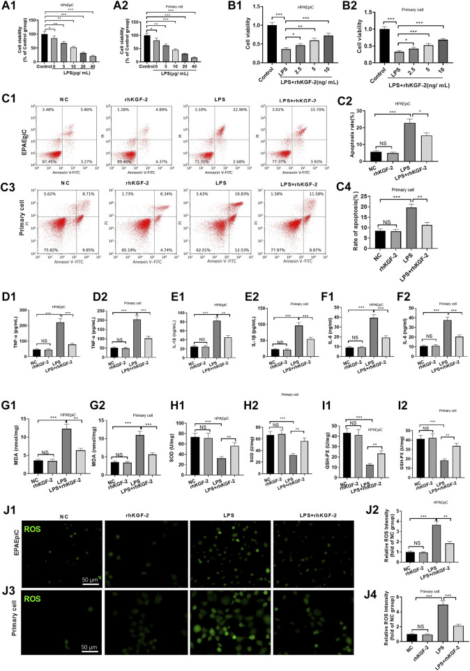
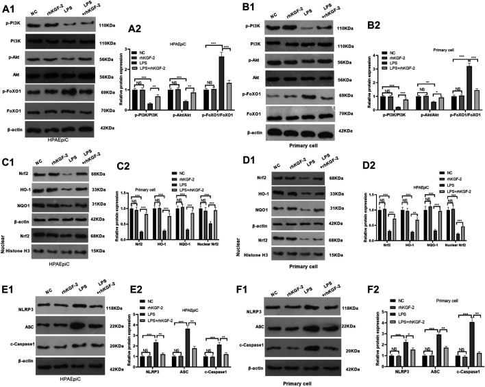
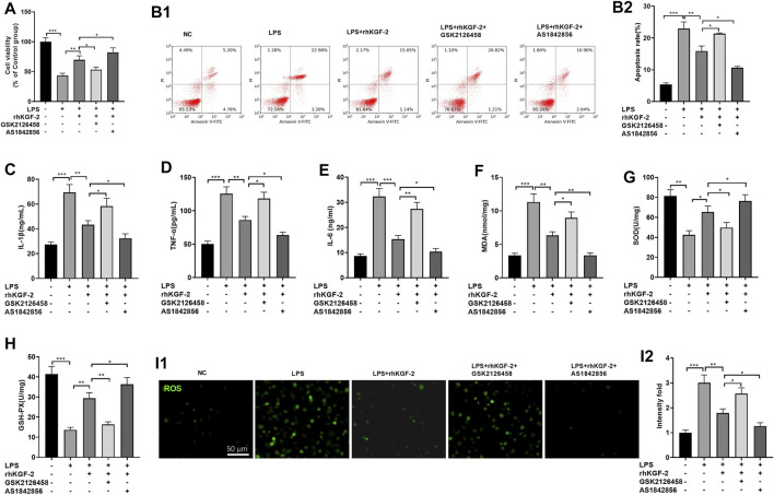
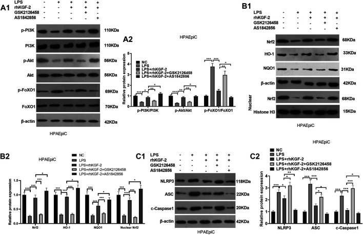
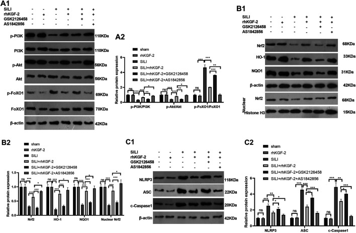
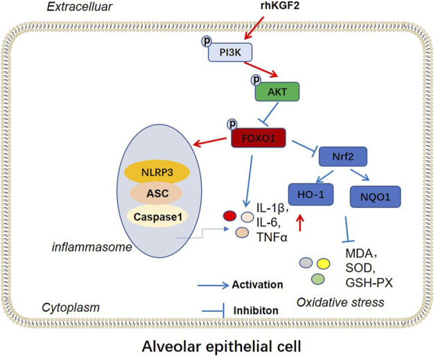
Smoke inhalation injury is an acute pathological change caused by thermal stimulation or toxic substance absorption through respiratory epithelial cells. This study aims to probe the protective effect and mechanism of recombinant human keratinocyte growth factor 2 (rhKGF-2) against smoke inhalation-induced lung injury (SILI) in rats. The SILI was induced in rats using a smoke exposure model, which were then treated with rhKGF-2. The rat blood was collected for blood-gas analysis, and the levels of inflammatory factors and oxidative stress markers in the plasma were measured. The rat lung tissues were collected. The pathological changes and cell apoptosis were determined by hematoxylin-eosin (HE) staining and TdT-mediated dUTP nick end labeling (TUNEL) assay, and the PI3K/Akt/Nrf2/HO-1/NQO1, and FoxO1-NLRP3 inflammasome expression were verified by western blot (WB). Both of the human alveolar epithelial cell (HPAEpiC) and primary rat alveolar epithelial cell were exposed to lipopolysaccharide (LPS) for making in-vitro alveolar epithelial cell injury model. After treatment with rhKGF-2, GSK2126458 (PI3K inhibitor) and AS1842856 (FoxO1 inhibitor), the cell viability, apoptosis, inflammation, oxidative stress, reactive oxygen species (ROS), PI3K/Akt/Nrf2, HO-1/NQO1, and FoxO1-NLRP3 in HPAEpiC and primary rat alveolar epithelial cell were examined. The data suggested that rhKGF-2 reduced LPS-induced HPAEpiC cell and primary rat alveolar epithelial cell apoptosis and the expression of inflammatory factors and oxidative stress factors. Moreover, rhKGF-2 improved the blood gas and alleviated SILI-induced lung histopathological injury in vivo via repressing inflammation, NLRP3 inflammasome activation and oxidative stress. Mechanistically, rhKGF-2 activated PI3K/Akt pathway, enhanced Nrf2/HO-1/NQO1 expression, and attenuated FoxO1-NLRP3 inflammasome both in vitro and in vivo. However, pharmaceutical inhibition of PI3K/Akt pathway attenuated rhKGF-2-mediated protective effects against SILI, while suppressing FoxO1 promoted rhKGF-2-mediated protective effects. Taken together, this study demonstrated that rhKGF-2 mitigated SILI by regulating the PI3K/Akt/Nrf2 pathway and the FoxO1-NLRP3 axis, which provides new reference in treating SILI.

Keywords: FoxO1; NLRP3; PI3K; inflammation; rhKGF-2; smoke inhalation injury.


Conflict of interest statement

The authors declare that the research was conducted in the absence of any commercial or financial relationships that could be construed as a potential conflict of interest.

Figures

FIGURE 1
FIGURE 1
The effect of rhKGF-2 on the cell viability, apoptosis, inflammation, oxidative and ROS generation of alveolar epithelial cells with LPS treatment. The HPAEpiC cells and primary rat alveolar epithelial cells were treated with LPS (5–20 μg/ml) and rhKGF-2 (2.5–10 ng/ml) for 24 h. A,B: Cell viability was determined by the MTT assay. The HPAEpiC cells and primary rat alveolar epithelial cells were treated with LPS (10 μg/ml) and rhKGF-2 (10 ng/ml) for 24 h. A1, A2, B1, B2: MTT assay were implemented to verify cell viability. The cell viability was calculated as the relative fold change of control group; C1–C4: FCM was conducted to analyze apoptosis. The rate of cell in the right upper quadrant was counted. D–O: The expression of TNF-α, IL-1β, IL-6, MDA, SOD, and GSH-PX in HPAEpiC cells and primary rat alveolar epithelial cells were examined by ELISA. The Y-axis shows the content of the detected mediators. p: The cellular ROS (marked by green color) level in HPAEpiC cells and primary rat alveolar epithelial cells was detected using the DCFDA/H2DCFDA-Cellular ROS Assay kit. The X-axis indicates the relative fluorescence intensity of ROS of NC group. Data are expressed as mean ± SD. All experiments were repeated for three times. Data difference was analyzed via ANOVA, and Student’s t test. NSP>0.05, *p<0.05, **p<0.01, ***p<0.001. N = 3.
FIGURE 2
FIGURE 2
The effect of rhKGF-2 on the expression of PI3K/Akt, Nrf2/HO-1/NQO1, and FoxO1-NLRP3-ASC-Caspase1 inflammasome. The HPAEpiC cells and primary rat alveolar epithelial cells were treated with LPS (10 μg/ml) and/or rhKGF-2 (10 ng/ml) for 24 h. A1-A2: WB was carried out to determine the expression of PI3K/Akt and FoxO1 in HPAEpiC cells. B1-B2: WB was carried out to determine the expression of PI3K/Akt and FoxO1 in primary rat alveolar epithelial cells. C1-C2. The protein levels of Nrf2, HO-1 and NQO1 in the HPAEpiC cells were detected by WB. D1-D2. The protein levels of Nrf2, HO-1 and NQO1 in the primary rat alveolar epithelial cells were detected by WB. E1-E2. WB was conducted to detect NLRP3-ASC-Caspase1 in HPAEpiC cells. F1-F2. WB was conducted to detect NLRP3-ASC-Caspase1 in primary rat alveolar epithelial cells. The p-PI3K, p-AKT and p-FoxO1 were calculated using PI3K, AKT and FoxO1 as internal control, respectively. β-actin was the internal control of Nrf2, HO-1, NQO1 and NLRP3-ASC-Caspase1. Nuclear Nrf2 was calculated using Histone H3 as internal control. The relative protein expression (Y-axis) was calculated as fold change of NC group. The X-axis showed the grouping names. Data are expressed as mean ± SD. All experiments were repeated for three times. Data difference was analyzed via ANOVA, and Student's t test. NSP>0.05, **p < 0.01, ***p < 0.001. N = 3.
FIGURE 3
FIGURE 3
The effect of PI3K inhibitor and FoxO1 inhibitor on rhKGF-2-mediated effect on HPAEpiC cells. LPS-treated HPAEpiC cells were adopted to construct a lung cell injury model, and then rhKGF-2 (10 ng/ml), GSK2126458 (0.5 nM) and AS1842856 (30 nM) were added for treatment for 24 h. (A): MTT assay was implemented to verify HPAEpiC viability. The cell viability was calculated as the relative fold change of control group. B1-B2. Flow cytometry was performed to verify HPAEpiC apoptosis. The rate of cell in the right upper quadrant was counted as apoptotic cells. C–H: ELISA was conducted to detect the expression of TNF-α (C), IL-1β (D), IL-6 (E), MDA (F), SOD (G), and GSH-PX. (H). The Y-axis shows the content of the detected mediators. (I). The cellular ROS level was detected using the DCFDA/H2DCFDA-Cellular ROS Assay kit. The X-axis indicates the relative fluorescence intensity of ROS of NC group. Data are expressed as mean ± SD. All experiments were repeated for three times. Data difference was analyzed via ANOVA, and Student's t test. *p<0.05, **p<0.01, ***p<0.001. N = 3.
FIGURE 4
FIGURE 4
The effect of PI3K inhibitor and FoxO1 inhibitor on the expression of PI3K/Akt, Nrf2/HO-1/NQO1, and FoxO1-NLRP3-ASC-Caspase1 inflammasome. BEAS-2B cells were treated with LPS (10 μg/ml), rhKGF-2 (10 ng/ml), GSK2126458 (0.5 nM) and/or AS1842856 (30 nM) for 24 h. A1-A2: WB was carried out to determine the expression of PI3K/Akt and FoxO1 in HPAEpiC cells. B1-B2. The protein levels of Nrf2, HO-1 and NQO1 in the HPAEpiC cells or nuclear were detected by WB. Nuclear Nrf2 was calculated using Histone H3 as internal control. C1-C2. WB was conducted to detect NLRP3-ASC-Caspase1 in HPAEpiC cells. The relative protein expression (Y-axis) was calculated as fold change of NC group. Data are expressed as mean ± SD. All experiments were repeated for three times. Data difference was analyzed via ANOVA, and Student's t test. *p<0.05, **p<0.01, ***p<0.001. N = 3.
FIGURE 5
FIGURE 5
The role of rhKGF-2 SILI rat model. The SILI rat model was established by inhalation of smoke. Then, rhKGF-2 (10 ng/kg body weight), GSK2126458 (300 μg/kg body weight) and AS1842856 (10 μg/kg body weight) were used for treating the SILI rats, the same volume of saline was administered on the rat in the sham group. (A–C). The arterial blood of rats was extracted to detect the values of PaO2, PaCO2, PaO2/FiO2 to analyze the pulmonary function. The Y-axis of panel A and B represents the partial pressure of O2 and CO2 (mmHg). The Y-axis of panel C represents the Oxygenation index (the ratio of arterial partial pressure of oxygen to inhaled oxygen concentration) (mmHg). (D). The wet weight (W) and dry weight of the rat lung were measured, and the W/D ratio was calculated to evaluate the pulmonary edema. (E). HE staining was performed to observe the pathological changes of rat lung in each group. (F). The pathological scores of rat lung in each group were counted. (G). TUNEL assay was implemented to analyze apoptosis in the lung tissues. (H). The number of TUNEL-positive cells in an area of 0.1 mm2 was calculated. Scale bar = 50 μm. Data are expressed as mean ± SD. All experiments were repeated for three times. Data difference was analyzed via ANOVA, and Student's t test. NSP>0.05, *p<0.05, **p<0.01, ***p<0.001. Five rats were involved in each group (N = 5).
FIGURE 6
FIGURE 6
rhKGF-2 eased the inflammation and oxidative stress in the SILI rat model. The SILI rat model was established by inhalation of smoke. Then, rhKGF-2 (10 ng/kg body weight), GSK2126458 (300 μg/kg body weight) and AS1842856 (10 μg/kg body weight) were used for were used for treating the SILI rats, the same volume of saline was administered on the rat in the sham group. A–F: The release of pro-inflammatory cytokines TNF-α (A), IL-1β (B), hs-CRP (C), and the levels of oxidative stress markers MDA (D), SOD (E), and GSH-PX. (F) in the lung of rats were determined by ELISA. The Y-axis shows the content of the detected mediators. The X-axis shows the grouping name. Data are expressed as mean ± SD. All experiments were repeated for three times. Data difference was analyzed via ANOVA, and Student’s t test. NSP>0.05, *p<0.05, **p<0.01, ***p<0.001. Five rats were involved in each group (N = 5).
FIGURE 7
FIGURE 7
rhKGF-2 has a role in regulating the activation of PI3K/Akt pathway, Nrf2/HO-1/NQO1 pathway and FoxO1-NLRP3-ASC-Caspase1 inflammasome in vivo. The SILI rat model was established by inhalation of smoke. Then, rhKGF-2 (10 ng/kg body weight), GSK2126458 (300 μg/kg body weight) and AS1842856 (10 μg/kg body weight) were used for were used for treating the SILI rats, the same volume of saline was administered on the rat in the sham group. Left lung tissues of rats in each group were collected for detection. A1-A2: WB was carried out to determine the expression of PI3K/Akt and FoxO1 in the lung tissues. B1-B2. The protein levels of Nrf2, HO-1 and NQO1 in the lung tissues. or nuclear were detected by WB. Nuclear Nrf2 was calculated using Histone H3 as internal control. C1-C2. WB was conducted to detect NLRP3-ASC-Caspase1 in the lung tissues. The relative protein expression (Y-axis) was calculated as fold change of sham group. Data are expressed as mean ± SD. All experiments were repeated for three times. Data difference was analyzed via ANOVA, and Student's t test. NSP>0.05, *p<0.05, **p<0.01, ***p<0.001. Five rats were involved in each group (N = 5).
FIGURE 8
FIGURE 8
The mechanism diagram. rhKGF2 enhanced the activation of PI3K/AKT pathway, inhibited the phosphorylation of FoxO1, thus repressing NLRP3-ASC-Caspase1 inflammasome and promoted Nrf2/HO-1/NQO1 axis. Finally, rhKGF2 repressed inflammation and oxidative stress of alveolar epithelial cells.

Similar articles

Cited by

References

    1. Arcidiacono B., Chiefari E., Messineo S., Bilotta F. L., Pastore I., Corigliano D. M., et al. (2018). HMGA1 Is a Novel Transcriptional Regulator of the FoxO1 Gene. Endocrine 60 (1), 56–64. 10.1007/s12020-017-1445-8 - DOI - PMC - PubMed
    1. Bi J., Tong L., Zhu X., Yang D., Bai C., Song Y., et al. (2014). Keratinocyte Growth Factor‐2 Intratracheal Instillation Significantly Attenuates Ventilator‐induced Lung Injury in Rats. J. Cel. Mol. Med. 18 (6), 1226–1235. 10.1111/jcmm.12269 - DOI - PMC - PubMed
    1. Chao C.-M., Yahya F., Moiseenko A., Tiozzo C., Shrestha A., Ahmadvand N., et al. (2017). Fgf10deficiency Is Causative for Lethality in a Mouse Model of Bronchopulmonary Dysplasia. J. Pathol. 241 (1), 91–103. 10.1002/path.4834 - DOI - PMC - PubMed
    1. Chen J., Wang Z. G., Zheng Z. M., Chen Y., Khor S., Shi K., et al. (2017). Neuron and Microglia/macrophage-Derived FGF10 Activate Neuronal FGFR2/PI3K/Akt Signaling and Inhibit Microglia/macrophages TLR4/NF-κb-dependent Neuroinflammation to Improve Functional Recovery after Spinal Cord Injury. Cell Death Dis 8 (10), e3090. 10.1038/cddis.2017.490 - DOI - PMC - PubMed
    1. Chen S., Li X., Wang Y., Mu P., Chen C., Huang P., et al. (2019). Ginsenoside Rb1 Attenuates Intestinal Ischemia/reperfusion-induced Inflammation and Oxidative Stress via Activation of the PI3K/Akt/Nrf2 Signaling Pathway. Mol. Med. Rep. 19 (5), 3633–3641. 10.3892/mmr.2019.10018 - DOI - PMC - PubMed