. 2021 Jul 23:12:685984.
doi: 10.3389/fimmu.2021.685984. eCollection 2021.

Exosome-Depleted Excretory-Secretory Products of the Fourth-Stage Larval Angiostrongylus cantonensis Promotes Alternative Activation of Macrophages Through Metabolic Reprogramming by the PI3K-Akt Pathway

Affiliations

Exosome-Depleted Excretory-Secretory Products of the Fourth-Stage Larval Angiostrongylus cantonensis Promotes Alternative Activation of Macrophages Through Metabolic Reprogramming by the PI3K-Akt Pathway

Shuo Wan et al. Front Immunol. .

Abstract

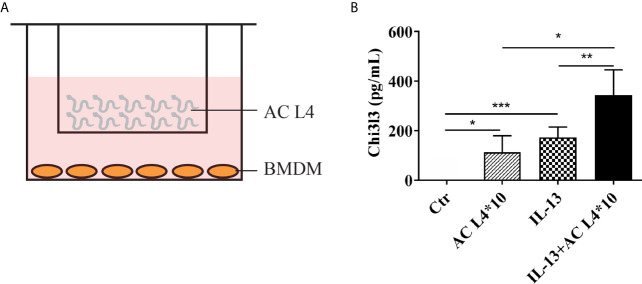
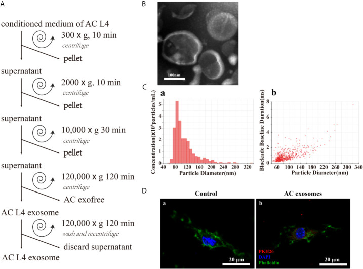
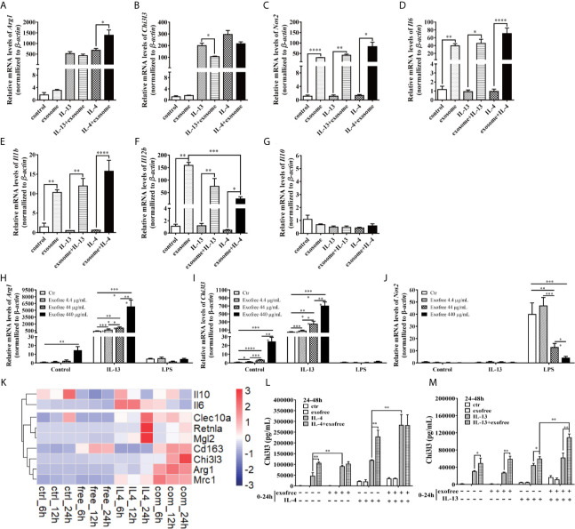
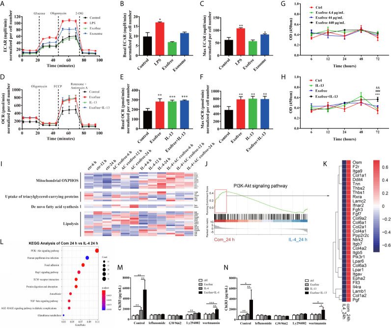
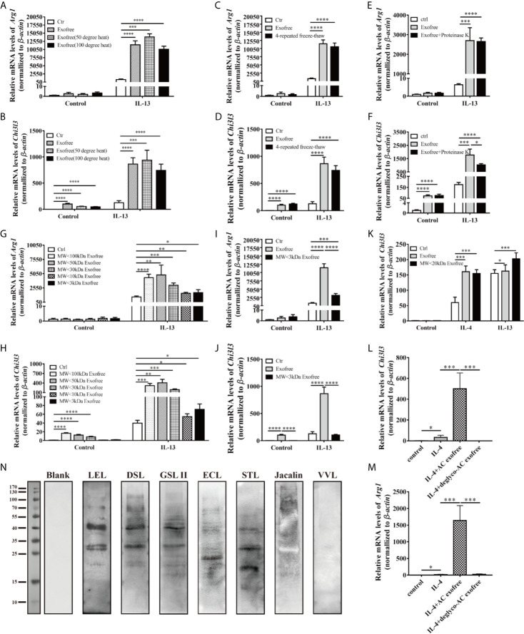
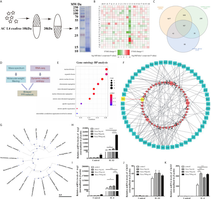
Angiostrongylus cantonensis (AC), which parasitizes in the brain of the non-permissive host, such as mouse and human, is an etiologic agent of eosinophilic meningitis. Excretory-secretory (ES) products play an important role in the interaction between parasites and hosts' immune responses. Inflammatory macrophages are responsible for eosinophilic meningitis induced by AC, and the soluble antigens of Angiostrongylus cantonensis fourth stage larva (AC L4), a mimic of dead AC L4, aggravate eosinophilic meningitis in AC-infected mice model via promoting alternative activation of macrophages. In this study, we investigated the key molecules in the ES products of AC L4 on macrophages and observed the relationship between metabolic reprogramming and the PI3K-Akt pathway. First, a co-culture system of macrophage and AC L4 was established to define the role of AC L4 ES products on macrophage polarization. Then, AC L4 exosome and exosome-depleted excretory-secretory products (exofree) were separated from AC L4 ES products using differential centrifugation, and their distinct roles on macrophage polarization were confirmed using qPCR and ELISA experiments. Moreover, AC L4 exofree induced alternative activation of macrophages, which is partially associated with metabolic reprogramming by the PI3K-Akt pathway. Next, lectin blot and deglycosylation assay were done, suggesting the key role of N-linked glycoproteins in exofree. Then, glycoproteomic analysis of exofree and RNA-seq analysis of exofree-treated macrophage were performed. Bi-layer PPI network analysis based on these results identified macrophage-related protein Hexa as a key molecule in inducing alternative activation of macrophages. Our results indicate a great value for research of helminth-derived immunoregulatory molecules, which might contribute to drug development for immune-related diseases.

Keywords: Angiostrongylus cantonensis; N-linked glycoproteins; exosome-depleted excretory-secretory products; macrophage polarization; mechanism.


Conflict of interest statement

The authors declare that the research was conducted in the absence of any commercial or financial relationships that could be construed as a potential conflict of interest.

Figures

Figure 1
Figure 1
Excretory-secretory products in AC L4 induce M2 polarization of macrophage. (A) Diagram of co-culture system with 10 AC L4 above the membrane and BMDMs below the membrane. (B) ELISA analysis of Chi3l3 of BMDMs in the presence of 10 AC L4, IL-13, and IL-13+10 AC L4 for 24 h. Data information: *P<0.05, **P<0.01.
Figure 2
Figure 2
Excretory-secretory products from the fourth stage larva of A. cantonensis could be further classified into AC L4 exosome and AC L4 exofree. (A) Scheme of AC L4 exosomes and AC L4 exofree isolation by differential ultracentrifugation from conditioned medium of the fourth stage larva of A. cantonensis. (B) Morphological characterization of AC L4 exosomes were identified using transmission electron microscope. (C) Concentration and size distribution of AC L4 exosomes were measured by IZON qNano particle analyzer. (D) BMDMs were incubated with PBS (a) or 5 μg/ml PKH26-labeled AC L4 exosomes (b) for 1 h, and a confocal analysis was performed to evaluate exosome internalization. Phalloidin was used to label actin (green), and DAPI was used for the staining of nuclei (blue). Scale bar=20 μm.
Figure 3
Figure 3
AC L4 exofree could promote a M2 polarization, while AC L4 exosomes induce M1 polarization of macrophage. (A–G) Macrophage activation markers Arg1 (A), Chi3l3 (B), Nos2 (C), and related interleukins Il6 (D), Il1b (E), Il12b (F), and Il10 (G) expression level were measured in the presence of AC L4 exosome, IL-4, IL-4+exosome, IL-13, and IL-13+exosome for 24 h using qPCR. The working concentration of AC L4 exosome is 25 μg/ml. The working concentration of IL-13 and IL-4 is 10 ng/ml. (H–J) Macrophage activation markers Arg1 (H), Chi3l3 (I), Nos2 (J) were measured in the presence of exofree, IL-4, IL-4+exofree, IL-13, and IL-13+exofree for 24 h using qPCR. (K) Signature gene expression profile of macrophage in the presence of PBS (ctr), free (AC L4 exofree), IL-4, com (IL-4+AC L4 exofree), and 6, 12, and 24 h represent 6, 12, and 24 h after the treatment, respectively. The working concentration of AC L4 exofree is 440 μg/ml. The working concentration of IL-13/IL-4 is 10 ng/ml. The working concentration of LPS is 0.05 μg/ml. The horizontal axis represents genes, and vertical coordinates represent types of treatment. (L, M) BMDMs were stimulated with PBS, AC L4 exofree, IL-4, IL-4+ AC L4 exofree, IL-13, IL-13+ AC L4 exofree in 0~24 h The culture medium was discarded. BMDMs were then washed with PBS and restimulated in 24~48 h as shown. And Chi3l3 protein level of 24~48 h culture medium was measured using ELISA. The working concentration of AC L4 exofree is 440 μg/ml. The working concentration of IL-13/IL-4 is 10 ng/ml. The detailed experiment information refer to Supplementary Table 2 . Data information: *P<0.05, **P<0.01, ***P<0.001, ****P<0.0001.
Figure 4
Figure 4
Metabolic Reprogramming by the PI3K-Akt pathway plays a vital role in AC L4 exofree–induced M2 polarization of macrophages. (A–C) BMDMs were cultured for 24 h in medium alone or treated with LPS, exofree, exosome, and then the ECAR (A) was monitored using the Seahorse Bioscience extracellular flux analyzer in real time. Dotted lines indicate incubation of cells with the indicated compounds. Basal ECAR (B) and Max ECAR (C) of BMDMs were calculated. *P<0.05, **P<0.01, compared with the control group. In this experiment, LPS concentration is 0.05 μg/ml, AC L4 exosome concentration is 25 μg/ml, and AC L4 exofree concentration is 44 μg/ml. (D–F) BMDMs were cultured for 24 h in medium alone or treated with exofree, IL-13, or exofree+IL-13, and then the OCR (D) was monitored using the Seahorse Bioscience extracellular flux analyzer in real time. Dotted lines indicate incubation of cells with the indicated compounds. Basal OCR (E) and Max ECAR (F) of BMDMs were calculated. **P<0.01, ***P<0.001, compared with the control group. In this experiment, IL-13 concentration is 10 ng/ml, and AC L4 exofree concentration is 44 μg/ml. (G, H) Proliferation of BMDMs was determined with CCK8 assay at 6, 12, 24, 48, 72 h after treatment of PBS and exofree (G). AC L4 exofree concentration in this experiment is 4.4, 44, 440 μg/ml. (H) Proliferation of BMDMs was determined with CCK8 assay at 6, 12, 24, 48, 72 h after treatment of PBS, IL-13, exofree, IL-13+exofree. In this experiment, AC L4 exofree concentration is 440 μg/ml, and IL-13 concentration is 10 ng/ml. &&P<0.01, compared with the control group; ###P<0.001, compared with the IL-13 group; ***P<0.001, compared with the exofree group. (I) Gene expression profile of BMDMs related to mitochondrial OXPHOS, nutrient uptake, de novo fatty acid synthesis, and lipolysis metabolism process were shown in a heatmap. BMDMs were treated with PBS (ctr), free (AC L4 exofree), IL-4, com (IL-4+AC L4 exofree) for 6, 12, and 24 h, respectively. The horizontal axis represents genes, and vertical coordinates represent types of treatment. In this experiment, AC L4 exofree concentration is 440 μg/ml, and IL-4 concentration is 10 ng/ml. (J) GSEA analysis of genes enriched in PI3K-Akt signaling pathway was performed using R package “clusterprofiler.” (K) Signature genes enriched in PI3K-Akt signaling pathway through GSEA analysis were shown in a heatmap. (L) GO enrichment analysis (biological process) of highly expressed genes in the presence of IL-4 and AC L4 exofree, compared with IL-4 treatment, was performed using R package “clusterprofiler.” (M, N) BMDMs were treated with PBS, exofree, IL-13, IL-13+exofree (M), and PBS, exofree, IL-4, IL-4+exofree (N) in the presence of leflunomide (a STAT6 inhibitor), GW9662 (a PPARγ antagonist), Ly294002 (PI3K inhibitor), and wortmannin (PI3K inhibitor) for 24 h. The protein levels of Chi3l3 in the culture medium were measured using ELISA. The following reagents were used in this experiment: Leflunomide (100 μM), Ly294002 (10 μM), Wortmannin (100 nM), GW9662 (1 μg/ml). *P<0.05, **P<0.01.
Figure 5
Figure 5
Chemical and physical property analysis of AC L4 exofree suggested glycoproteins as the key components in AC L4 exofree–induced M2 polarization of macrophage. (A, B) qPCR analysis of Arg1 (A), Chi3l3 (B) mRNA level of BMDMs in the presence of PBS, AC L4 exofree, AC L4 exofree (50°C water bath, 15min), AC L4 exofree (100°C water bath, 15min), IL-13, AC L4 exofree+IL-13, AC L4 exofree (50°C water bath, 15min)+IL-13, AC L4 exofree (100°C water bath, 15 min)+IL-13 at 24 h was performed. (C, D) qPCR analysis of Arg1 (C), Chi3l3 (D) mRNA level of BMDMs in the presence of PBS, AC L4 exofree, AC L4 exofree (repeated freeze-thaw cycles for four times) at 24 h was performed. (E, F) qPCR analysis of Arg1 (E), Chi3l3 (F) mRNA level of BMDMs in the presence of PBS, AC L4 exofree, AC L4 exofree (100 μg/ml proteinase K treatment: 58°C water bath, 2 h; proteinase K inactivation: 100°C water bath, 15 min) at 24 h was performed. (G, H) qPCR analysis of Arg1 (G), Chi3l3 (H) mRNA level of BMDMs in the presence of PBS, AC L4 exofree, AC L4 exofree (<3 kDa fraction), AC L4 exofree (<10 kDa fraction), AC L4 exofree (<30 kDa fraction), AC L4 exofree (<50 kDa fraction), AC L4 exofree (<100 kDa fraction), IL-13, AC L4 exofree+IL-13, AC L4 exofree (<3 kDa fraction)+IL-13, AC L4 exofree (<10 kDa fraction)+IL-13, AC L4 exofree (<30 kDa fraction)+IL-13, AC L4 exofree (<50 kDa fraction)+IL-13, AC L4 exofree (<100 kDa fraction)+IL-13, at 24 h was performed. (I, J) qPCR analysis of Arg1 (I) and Chi3l3 (J) mRNA level of BMDMs in the presence of PBS, AC L4 exofree, AC L4 exofree (<3 kDa fraction), IL-13, AC L4 exofree+IL-13, AC L4 exofree (<3 kDa fraction)+IL-13 at 24 h was performed. In (A–J), the concentration of exofree is 440 μg/ml and the concentration of IL-13 is 10 ng/ml. (K) qPCR analysis of Chi3l3 mRNA level of BMDMs in the presence of PBS, AC L4 exofree, AC L4 exofree (<20 kDa fraction), IL-13, AC L4 exofree+IL-13, AC L4 exofree (<20 kDa fraction)+IL-13 at 24 h was performed. In (K), the concentration of exofree is 44 μg/ml, the concentration of IL-13 is 10 ng/ml. (L, M) Control (PBS) or AC L4 exofree or deglycosylated AC L4 exofree were used to stimulate BMDMs. And mRNA expression level of Chi3l3 (L) and Arg1 (M) were measured 24 h after the stimulation using qPCR. (N) Glycoproteins in AC L4 exofree were measured by lectin blot. Membranes were incubated with PBS (lane Blank), lectin LEL, DSL, GSL II, ECL, STL, Jacalin, and VVL as primary antibody, respectively. The same amount of exofree is used in experiments from (A–M). Data information: *P<0.05, **P<0.01, ***P<0.001, ****P<0.0001.
Figure 6
Figure 6
AC-derived Hexa act as a key ingredient in promoting M2 activation of macrophages. (A) Diagram of isolation of 10~20 kDa-AC L4 exofree using ultrafiltration tubes, and the efficiency was confirmed using western blot. (B) Heatmap showed the relative frequencies of amino acids between the asparagine (Asn) position of N-linked glycoproteins in AC L4 exofree. Enrichment level of the amino acid at indicate position is shown in different colors. Red means high enrichment level, and green means low enrichment level. (C) Venn diagram of AC L4 exosome, AC L4 exofree proteins identified using mass spectrum analysis. (D) Diagram of Bi-layer PPI network analysis using AC L4 exofree proteins and RNA-seq data of BMDMs. (E) GO enrichment analysis (Biological process) of exofree-induced TCGs of BMDMs ( Supplementary Figure 5B ). (F) A protein-gene network was constructed for worm proteins with molecular weight less than 50kDa and TCGs of exofree-, IL-4-, and exofree+IL-4-treated BMDMs ( Supplementary Figure 5A ), based on the LC-MS/MS and RNA-seq data. (G) Phylogene tree of Hexa protein in 17 species. (H, J) qPCR analysis of mRNA level of Arg1 (H) and Chi3l3 (J) were measured in the presence of PBS, Hexa 5 ng/ml, Hexa 50 ng/ml, Hexa 500 ng/ml, IL-13 10 ng/ml, IL-13+Hexa 5 ng/ml, IL-13+Hexa 50 ng/ml, IL-13+Hexa 500 ng/ml for 24 h. (I, K) qPCR analysis of mRNA level of Arg1 (I) and Chi3l3 (K) were measured in the presence of PBS, Hexa 5 ng/ml, Hexa 50 ng/ml, Hexa 500 ng/ml, IL-4 10 ng/ml, IL-4+Hexa 5 ng/ml, IL-4+Hexa 50 ng/ml, IL-4+Hexa 500 ng/ml for 24 h. Data information: *P<0.05, ***P<0.001, ****P<0.0001.

Similar articles

Cited by

References

    1. Zhao J, Lv Z, Wang F, Wei J, Zhang Q, Li S, et al. . Ym1, an Eosinophilic Chemotactic Factor, Participates in the Brain Inflammation Induced by Angiostrongylus Cantonensis in Mice. Parasitol Res (2013) 112:2689–95. 10.1007/s00436-013-3436-x - DOI - PubMed
    1. Wan S, Sun X, Wu F, Yu Z, Wang L, Lin D, et al. . Chi3l3: A Potential Key Orchestrator of Eosinophil Recruitment in Meningitis Induced by Angiostrongylus Cantonensis. J Neuroinflamm (2018) 15:31. 10.1186/s12974-018-1071-2 - DOI - PMC - PubMed
    1. Rodriguez E, Noya V, Cervi L, Chiribao ML, Brossard N, Chiale C, et al. . Glycans From Fasciola Hepatica Modulate the Host Immune Response and TLR-Induced Maturation of Dendritic Cells. PloS Negl Trop Dis (2015) 9:e0004234. 10.1371/journal.pntd.0004234 - DOI - PMC - PubMed
    1. Prasanphanich NS, Luyai AE, Song X, Heimburg-Molinaro J, Mandalasi M, Mickum M, et al. . Immunization With Recombinantly Expressed Glycan Antigens From Schistosoma Mansoni Induces Glycan-Specific Antibodies Against the Parasite. Glycobiology (2014) 24:619–37. 10.1093/glycob/cwu027 - DOI - PMC - PubMed
    1. Eichenberger RM, Talukder MH, Field MA, Wangchuk P, Giacomin P, Loukas A, et al. . Characterization of Trichuris Muris Secreted Proteins and Extracellular Vesicles Provides New Insights Into Host-Parasite Communication. J Extracell Vesicles (2018) 7:1428004. 10.1080/20013078.2018.1428004 - DOI - PMC - PubMed

Publication types

MeSH terms

Substances