. 2021 Jul 23:12:709695.
doi: 10.3389/fimmu.2021.709695. eCollection 2021.

Identification of Disease-Associated Cryptococcal Proteins Reactive With Serum IgG From Cryptococcal Meningitis Patients

Affiliations

Identification of Disease-Associated Cryptococcal Proteins Reactive With Serum IgG From Cryptococcal Meningitis Patients

A Elisabeth Gressler et al. Front Immunol. .

Abstract

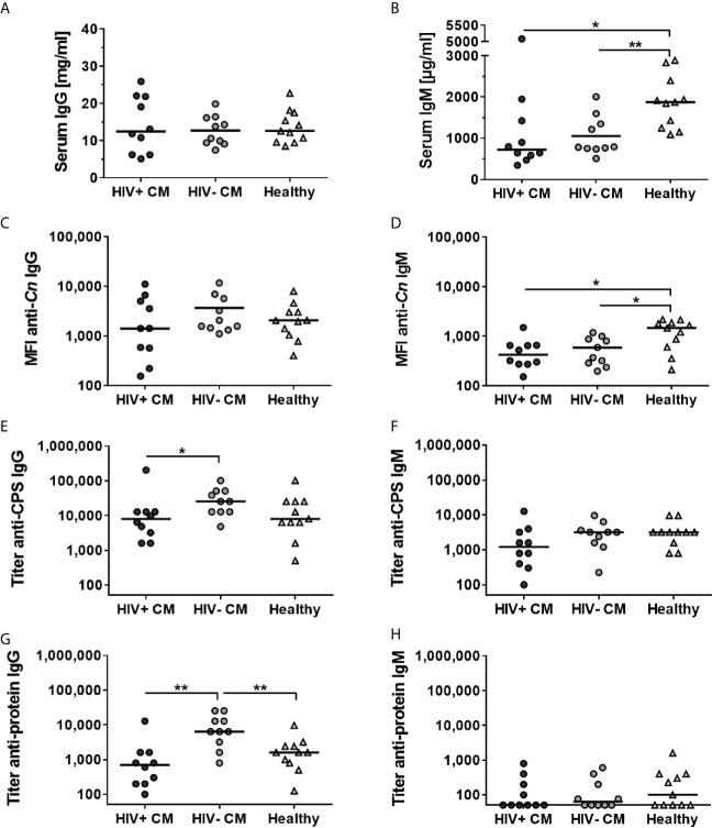
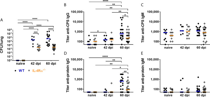
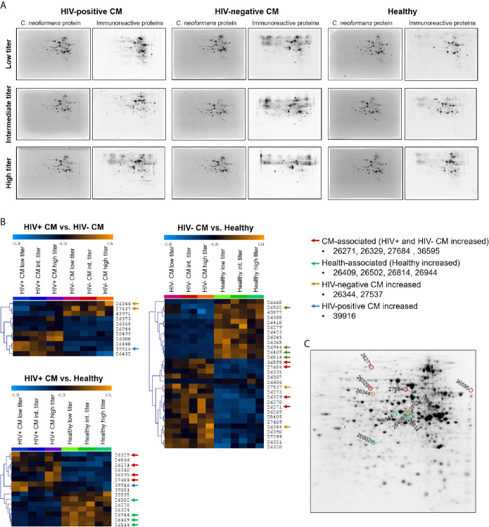
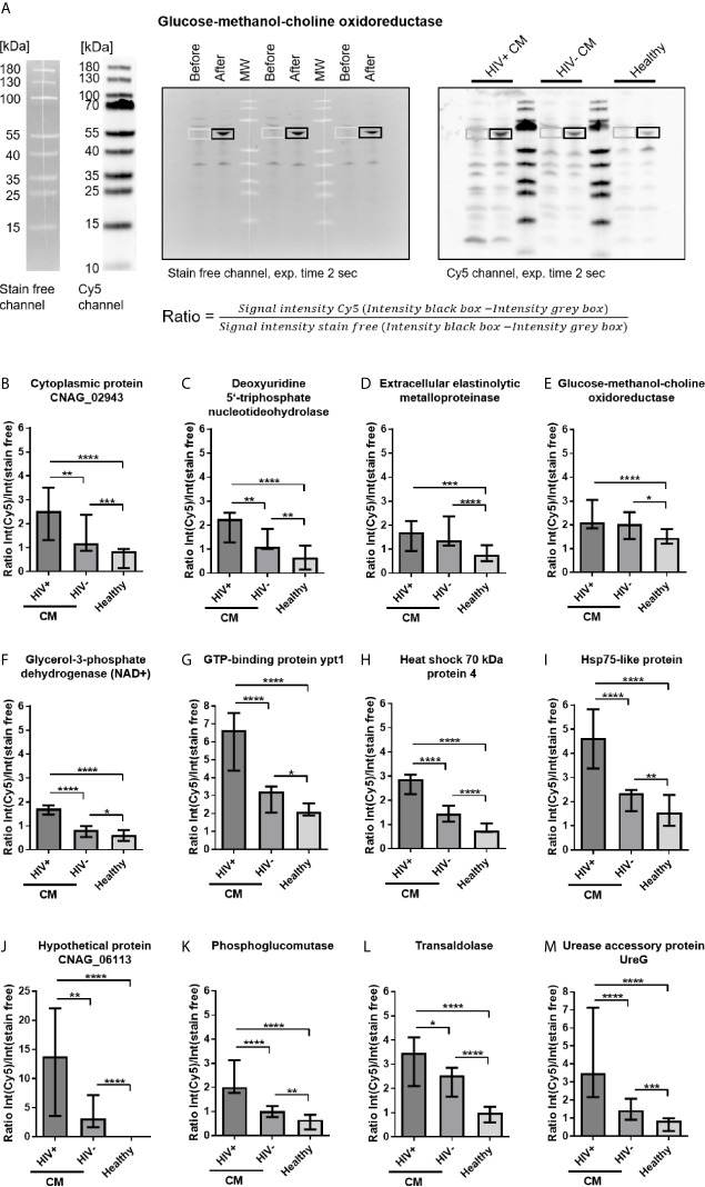
Cryptococcus neoformans, an opportunistic fungal pathogen ubiquitously present in the environment, causes cryptococcal meningitis (CM) mainly in immunocompromised patients, such as AIDS patients. We aimed to identify disease-associated cryptococcal protein antigens targeted by the human humoral immune response. Therefore, we used sera from Colombian CM patients, with or without HIV infection, and from healthy individuals living in the same region. Serological analysis revealed increased titers of anti-cryptococcal IgG in HIV-negative CM patients, but not HIV-positive CM patients, compared to healthy controls. In contrast, titers of anti-cryptococcal IgM were not affected by CM. Furthermore, we detected pre-existing IgG and IgM antibodies even in sera from healthy individuals. The observed induction of anti-cryptococcal IgG but not IgM during CM was supported by analysis of sera from C. neoformans-infected mice. Stronger increase in IgG was found in wild type mice with high lung fungal burden compared to IL-4Rα-deficient mice showing low lung fungal burden. To identify the proteins targeted by human anti-cryptococcal IgG antibodies, we applied a quantitative 2D immunoproteome approach identifying cryptococcal protein spots preferentially recognized by sera from CM patients or healthy individuals followed by mass spectrometry analysis. Twenty-three cryptococcal proteins were recombinantly expressed and confirmed to be immunoreactive with human sera. Fourteen of them were newly described as immunoreactive proteins. Twelve proteins were classified as disease-associated antigens, based on significantly stronger immunoreactivity with sera from CM patients compared to healthy individuals. The proteins identified in our screen significantly expand the pool of cryptococcal proteins with potential for (i) development of novel anti-cryptococcal agents based on implications in cryptococcal virulence or survival, or (ii) development of an anti-cryptococcal vaccine, as several candidates lack homology to human proteins and are localized extracellularly. Furthermore, this study defines pre-existing anti-cryptococcal immunoreactivity in healthy individuals at a molecular level, identifying target antigens recognized by sera from healthy control persons.

Keywords: Cryptococcus neoformans; cryptococcal meningitis; fungal infection; human samples; humoral immunity; immunoproteomics.


Conflict of interest statement

The authors declare that the research was conducted in the absence of any commercial or financial relationships that could be construed as a potential conflict of interest.

Figures

Figure 1
Figure 1
Total and Cryptococcus neoformans-specific IgG and IgM antibodies differ between quenchable sera from cryptococcal meningitis patients and healthy controls. Human sera from patients with cryptococcal meningitis (CM), either HIV-positive (HIV+) or HIV-negative (HIV-), and healthy control persons were analyzed. Concentration of (A) total serum IgG antibodies was similar in all groups, whereas concentration of (B) total serum IgM antibodies was increased in the healthy Colombian control group. C. neoformans-specific antibodies directed against intact cryptococcal cells (anti-Cn) (C) IgG or (D) IgM antibodies were quantified using flow cytometry. Median fluorescent intensity (MFI) was similar for anti-Cn IgG antibodies in all groups, but significantly enhanced for anti-Cn IgM antibodies in healthy control persons. Titers of (E) IgG and (F) IgM directed capsular polysaccharides (CPS) as well as titers of (G) IgG and (H) IgM directed against cryptococcal proteins were determined using ELISA. Titers of anti-CPS IgG and anti-protein IgG were increased in HIV-negative CM patients compared to HIV-positive CM patients and healthy control persons, whereas titers of anti-CPS IgM and anti-protein IgM were similar between all groups. Each dot represents an individual serum and lines indicate median values. Statistical analysis was carried out using the Mann–Whitney U test for comparison of two groups. Asterisks indicate significant difference.
Figure 2
Figure 2
Lung fungal burden and Cryptococcus neoformans-specific antibody titers in wild type (WT) and IL-4Rα-deficient (IL-4Rα-/-) mice. WT and IL-4Rα-/- mice were infected intranasally with 500 colony forming units (CFU) of Cryptococcus neoformans serotype D strain 1841. (A) Lung fungal burdens were determined in non-infected (naïve) mice, on 42 days post infection (dpi) and 60 dpi. Fungal burden were significantly higher on both 42 and 60 dpi in WT mice compared to IL-4Rα-/- mice. Titers of IgG and IgM antibodies directed against capsular polysaccharides (CPS) and cryptococcal proteins were determined using ELISA analysis. Titers of (B) anti-CPS IgG and (D) anti-protein IgG increased upon infection for both genotypes, but were significantly higher in WT compared to IL−4Rα−/− mice on 60 dpi. In contrast, titers of (C) anti-CPS IgM and (E) anti-protein IgM were unaffected by the progression of infection, independently of the genotype. Sera from seven to 23 mice from at least two independent experiments were analyzed per genotype. Each dot represents an individual serum and lines indicate median values. Statistical analysis was carried out using the Mann–Whitney U test for comparison of two groups. Asterisks indicate significant difference.
Figure 3
Figure 3
Two-dimensional (2D) analysis of the immunoproteome recognized by cryptococcal meningitis (CM) patients and healthy individuals revealed infection- and health-associated spots. Cryptococcus neoformans proteins (strain H99) were separated by 2D gel electrophoresis and transferred onto nitrocellulose membranes. (A) Representative blot images of total cryptococcal proteins (stained with UV-activated 2,2,2-trichloroethanol (TCE)) and immunoreactive protein spots bound by serum IgG (detected using AF-647 labeled secondary antibody) are shown. The contrast and brightness of the images were adjusted for publication and do not reflect actual signal intensities. (B) Heat maps of immunoreactive protein spots’ fluorescence signals recognized with different intensities by sera from different groups (p < 0.01). Spots were either (i) CM-associated (red arrows) – significantly stronger reactivity with CM patient sera (HIV-positive and HIV-negative) compared to healthy individuals, (ii) health-associated (green arrows) – significantly stronger reactivity with sera from healthy individuals compared to CM patients (HIV-positive and HIV-negative). Three spots were found that showed higher reactivity of (iii) HIV-negative sera (yellow arrows) or (iv) HIV-positive sera (blue arrows) compared to the respective other two groups. Immunoreactivity per sub-pool was determined in duplicates. Spot identifiers (row numbers) were automatically created by Delta 2D software. Analysis parameters: Test design: between-subjects, used Welch approximation, alpha: 0.01, p-values based on permutation, all permutations used: true, number of permutations per spot: 924, significance determined by standard Bonferroni correction, HCL: complete linkage, Euclidean Distance. (C) Spots of interest were highlighted in a 2D gel image. Proteins were stained with UV-activated TCE. Int., Intermediate.
Figure 4
Figure 4
Cryptococcal meningits (CM)-associated recombinant cryptococcal proteins reactive with serum IgG. (A) Escherichia coli lysates containing recombinant cryptococcal proteins were separated by SDS-PAGE. Samples before induction of recombinant protein expression and samples after induction of protein expression using IPTG were loaded, separated by MW markers, as representatively shown for glucose-methanol-choline oxidoreductase. E. coli proteins were blotted onto nitrocellulose membranes and (left) stained with 2,2,2-trichloroethanol (TCE, detection in stain free channel). Membranes were incubated with pooled sera from HIV-positive (HIV+) CM patients, HIV-negative (HIV-) CM patients, or healthy control persons, as marked in the blot. (Right) Proteins immunoreactive with human IgG were detected using polyclonal goat-anti human IgG coupled to Alexa Fluor 647 (AF-647, detected in Cy5 channel). Ratios of fluorescence intensities of the respective area in both channels were calculated to normalize the immunosignal (Cy5 channel) onto the amount of protein loaded (stain free channel). (B–M) CM-associated cryptococcal proteins reactive with serum IgG are shown. Proteins depicted showed significantly stronger reactivity with sera from CM patients (HIV+ and HIV-) compared to sera from healthy individuals. Median values and range of three independent experiments, comprising values from three different exposure times for each experiment, are shown. Statistical analysis was carried out using the Mann–Whitney U test for comparison of two groups. Asterisks indicate significant difference. MW, Molecular weight marker; exp., exposure.

Similar articles

Cited by

References

    1. Kwon-Chung KJ, Fraser JA, Doering TL, Wang Z, Janbon G, Idnurm A, et al. . Cryptococcus neoformans and Cryptococcus gattii, the Etiologic Agents of Cryptococcosis. Cold Spring Harb Perspect Med (2014) 4:1–27. 10.1101/cshperspect.a019760 - DOI - PMC - PubMed
    1. Negroni R, Arechavala AI. Pathogenesis of Cryptococcosis in Humans. In: Singh SK, editor. Human Emerging and Re-emerging Infections: Bacterial & Mycotic Infections. Hoboken, New Jersey, United States: John Wiley & Sons; (2016). p. 915–27. 10.1002/9781118644843.ch49 - DOI
    1. Soltani M, Bayat M, Hashemi SJ, Zia M, Pestechian N. Isolation of Cryptococcus neoformans and Other Opportunistic Fungi From Pigeon Droppings. J Res Med Sci (2013) 18:56–60. - PMC - PubMed
    1. Zarrin M, Jorfi M, Amirrajab N, Rostami M. Isolation of Cryptococcus neoformans From Pigeon Droppings in Ahwaz, Iran. Turk J Med Sci (2010) 40:313–6. 10.3906/sag-0808-10 - DOI
    1. Brito-Santos F, Trilles L, Firacative C, Wanke B, Carvalho-Costa FA, Nishikawa MM, et al. . Indoor Dust as a Source of Virulent Strains of the Agents of Cryptococcosis in the Rio Negro Micro-Region of the Brazilian Amazon. Microorganisms (2020) 8:682. 10.3390/microorganisms8050682 - DOI - PMC - PubMed

Publication types

MeSH terms

LinkOut - more resources