. 2021 Jul 2:12:692847.
doi: 10.3389/fpls.2021.692847. eCollection 2021.

Functional Relationship of Arabidopsis AOXs and PTOX Revealed via Transgenic Analysis

Affiliations

Functional Relationship of Arabidopsis AOXs and PTOX Revealed via Transgenic Analysis

Danfeng Wang et al. Front Plant Sci. .

Abstract

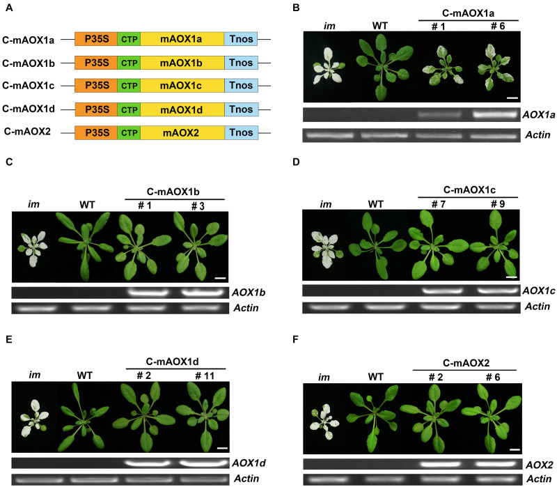
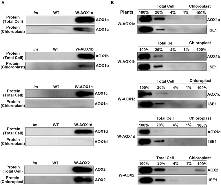
Alternative oxidase (AOX) and plastid terminal oxidase (PTOX) are terminal oxidases of electron transfer in mitochondria and chloroplasts, respectively. Here, taking advantage of the variegation phenotype of the Arabidopsis PTOX deficient mutant (im), we examined the functional relationship between PTOX and its five distantly related homologs (AOX1a, 1b, 1c, 1d, and AOX2). When engineered into chloroplasts, AOX1b, 1c, 1d, and AOX2 rescued the im defect, while AOX1a partially suppressed the mutant phenotype, indicating that AOXs could function as PQH2 oxidases. When the full length AOXs were overexpressed in im, only AOX1b and AOX2 rescued its variegation phenotype. In vivo fluorescence analysis of GFP-tagged AOXs and subcellular fractionation assays showed that AOX1b and AOX2 could partially enter chloroplasts while AOX1c and AOX1d were exclusively present in mitochondria. Surprisingly, the subcellular fractionation, but not the fluorescence analysis of GFP-tagged AOX1a, revealed that a small portion of AOX1a could sort into chloroplasts. We further fused and expressed the targeting peptides of AOXs with the mature form of PTOX in im individually; and found that targeting peptides of AOX1a, AOX1b, and AOX2, but not that of AOX1c or AOX1d, could direct PTOX into chloroplasts. It demonstrated that chloroplast-localized AOXs, but not mitochondria-localized AOXs, can functionally compensate for the PTOX deficiency in chloroplasts, providing a direct evidence for the functional relevance of AOX and PTOX, shedding light on the interaction between mitochondria and chloroplasts and the complex mechanisms of protein dual targeting in plant cells.

Keywords: alternative oxidase (AOX); chloroplasts; mitochondria; plastid terminal oxidase (PTOX); protein dual targeting; targeting peptide.


Conflict of interest statement

The authors declare that the research was conducted in the absence of any commercial or financial relationships that could be construed as a potential conflict of interest.

Figures

FIGURE 1
FIGURE 1
Expression of C-mAOX constructs in im. (A) Schematic diagram of C-mAOX1a, C-mAOX1b, C-mAOX1c, C-mAOX1d, and C-mAOX2. P35S, Cauliflower Mosaic Virus 35S promoter; Tnos, terminator of nos (nopaline synthetase); CTP, chloroplast targeting sequence from Arabidopsis thaliana RbcS1A (Rubisco small subunit); mAOX1a, mAOX1b, mAOX1c, mAOX1d, and mAOX2 are the mature proteins (the full length AOX minus the targeting peptide). (B–F) Phenotypes of im mutant, WT, and transgenic im mutants carrying 35S-driven C-mAOX1a, C-mAOX1b, C-mAOX1c, C-mAOX1d, and C-mAOX2. Expression of C-mAOX in im was examined at the RNA level by RT-PCR. All plants were grown for 4 weeks with a photoperiod of 16 h light/8 h dark cycle under light (∼100 μmol⋅m–2s–1) after initial low light (∼20 μmol⋅m–2s–1) growth for 5 days as described in the “Materials and Methods” section. Actin was used as quantity control. Bars = 1 cm.
FIGURE 2
FIGURE 2
Subcellular localization of C-mAOX1a, C-mAOX1b, C-mAOX1c, C-mAOX1d, and C-mAOX2. C-mAOX1a, C-mAOX1b, C-mAOX1c, C-mAOX1d, and C-mAOX2 tagged with a C-terminal GFP were transiently expressed under the control of 35S promoter in N. benthamiana leaves and observed by confocal microscopy. In each case, images of chlorophyll autofluorescence (Chl), GFP fluorescence (GFP), and merged chlorophyll and GFP fluorescence with bright-field (Merge) are shown. Scale bar = 20 μm.
FIGURE 3
FIGURE 3
Expression of W-AOX constructs in im. (A) Schematic diagram of W-AOX1a, W-AOX1b, W-AOX1c, W-AOX1d, and W-AOX2 constructs. P35S, Cauliflower Mosaic Virus 35S promoter; Tnos, terminator of nos (nopaline synthetase); WAOX1a, WAOX1b, WAOX1c, WAOX1d, and WAOX2 are the full length coding sequences. (B–F) Phenotypes of im mutant, WT, and three independent lines of transgenic im mutants carrying 35S-driven W-AOX1a, W-AOX1b, W-AOX1c, W-AOX1d, and W-AOX2. All plants were grown for 4 weeks with a photoperiod of 16 h light/8 h dark cycle under light (∼100 μmol⋅m–2s–1) after initial low light (∼20 μmol⋅m–2s–1) growth for 5 days as described in the “Materials and Methods” section. Total cell proteins was isolated from 10 mg of fresh leaf tissue, and subjected to 12% SDS-PAGE and probed with the corresponding AOX antibodies. Bars = 1 cm.
FIGURE 4
FIGURE 4
Subcellular localization of W-AOX1a, W-AOX1b, W-AOX1c, W-AOX1d, and W-AOX2. W-AOX1a, W-AOX1b, W-AOX1c, W-AOX1d, and W-AOX2 tagged with a C-terminal GFP were transiently expressed under the control of 35S promoter in N. benthamiana leaves and observed by confocal microscopy. (A) In each case, images of mitochondrial Mcherry fluorescence (Mt-Mcherry), GFP fluorescence (GFP), and merged Mcherry and GFP fluorescence with bright-field (Merge) are shown. Scale bar = 5 μm. (B) In each case, images of chlorophyll autofluorescence (Chl), GFP fluorescence (GFP), and merged chlorophyll and GFP fluorescence with bright-field (Merge) are shown. Scale bar = 20 μm.
FIGURE 5
FIGURE 5
W-AOX1a, W-AOX1b, and W-AOX2 are present in chloroplasts of the overexpression lines. (A) Immunoblotting analyses. Rosette leaves from 4-week-old plants as in Figure 3 served as the source of total cell proteins and of chloroplast membranes from lysed, Percoll gradient-purified chloroplasts (designated chloroplasts). Equal protein amounts were electrophoresed through 12% SDS polyacrylamide gels, and immunoblotting analyses were performed with the corresponding AOX antibodies. (B) Total cell proteins or proteins from gradient purified chloroplasts were isolated from the rosette leaves of 4-week-old W-AOX1a, W-AOX1b, W-AOX1c, W-AOX1d, and W-AOX2 plants. Samples containing equal chlorophyll amounts (2 μg chlorophyll) were electrophoresed through 12% SDS polyacrylamide gels, and immunoblotting analyses were performed with antibodies against the corresponding AOX, and ISE1, a highly expressed mitochondrial-specific protein (Stonebloom et al., 2009). The gels contained a dilution series (100, 20, 4, and 1%) of total cell proteins.
FIGURE 6
FIGURE 6
Expression of AOXTP-mPTOX constructs in im. (A) Schematic diagram of ATG-mPTOX, AOX1aTP-mPTOX, AOX1bTP-mPTOX, AOX1cTP-mPTOX, AOX1dTP-mPTOX, and AOX2TP-mPTOX constructs. P35S, Cauliflower Mosaic Virus 35S promoter; Tnos, terminator of nos (nopaline synthetase); ATG, initiation codon; 1aTP, AOX1a targeting sequence; 1bTP, AOX1b targeting sequence; 1cTP, AOX1c targeting sequence; 1dTP, AOX1d targeting sequence; 2TP, AOX2 targeting sequence. mPTOX, mature peptide sequence of PTOX. (B–G) Phenotypes of im mutant, WT, and three independent lines of transgenic im mutants carrying 35S-driven ATG-mPTOX, AOX1aTP-mPTOX, AOX1bTP-mPTOX, AOX1cTP-mPTOX, AOX1dTP-mPTOX, and AOX2TP-mPTOX. All plants were grown for 4 weeks with a photoperiod of 16 h light/8 h dark cycle under light (∼100 μmol⋅m–2s–1) after initial low light (∼20 μmol⋅m–2s–1) growth for 5 days as described in the “Materials and Methods” section. Total cell proteins were isolated from 10 mg fresh weight of leaf tissue (Total Cell). Chloroplast proteins were isolated from pellets obtained by centrifugation of lysed, Percoll gradient-purified chloroplasts (corresponding to 2 μg of chlorophyll). The protein samples were subjected to 12% SDS-PAGE and probed with PTOX antibody. Asterisk (*) marks unspecific bands detected by an antibody against PTOX. Bars = 1 cm.
FIGURE 7
FIGURE 7
Chlorophyll a fluorescence measurements. Chlorophyll fluorescence parameters were measured on intact leaves from W-AOX1b (A) and W-AOX2 (B) and wild-type and im grown for 7 weeks in soil under the conditions as described in the “Materials and Methods” section. The parameters included the following: Fv’/Fm’, the maximum efficiency of PSII photochemistry under different photo flux densities (PFD); ΦPSII, the quantum efficiency of PSII photochemistry at different photo flux densities; 1-qP, the redox state of the QA electron acceptor of PSII (Maxwell and Johnson, 2000; Müller et al., 2001).
FIGURE 8
FIGURE 8
NPQ analyses. Steady state light response NPQ (A) and rapid induction and dark relaxation NPQ kinetics (B) were measured on intact leaves from wild-type, im, W-AOX1b, and W-AOX2 seedlings grown for 7 weeks in soil under low light (20 μmol⋅m–2s–1) for 5 days at 16-h light/8-h dark daylight cycle, and then were transferred to the normal growth condition (23°C, 16-h light/8-h dark daylight cycle, 100 μmol⋅m–2s–1) as described in the “Materials and Methods” section. The data represent the average ± SD of four independent experiments.

References

    1. Abdallah F., Salamini F., Leister D. (2000). A prediction of size and evolutionary origin of the proteome of chloroplast of Arabidopsis. Trends Plan Sci. 5 141–142. 10.1016/s1360-1385(00)01574-0 - DOI - PubMed
    1. Baudisch B., Langner U., Garz I., Klösgen R. B. (2014). The exception proves the rule? Dual targeting of nuclear-encoded proteins into endosymbiotic organelles. New Phytol. 201 80–90. 10.1111/nph.12482 - DOI - PubMed
    1. Bennoun P. (2002). The present model for chlororespiration. Photosynth. Res. 73 273–277. 10.1023/A:1020479920622 - DOI - PubMed
    1. Bhuiyan N. H., Friso G., Poliakov A., Ponnala L., van Wijk K. J. (2015). MET1 is a thylakoid-associated TPR protein involved in photosystem II supercomplex formation and repair in Arabidopsis. Plant Cell 27 262–285. 10.1105/tpc.114.132787 - DOI - PMC - PubMed
    1. Blanco N. E., Guinea-Díaz M., Whelan J., Strand Å. (2014). Interaction between plastid and mitochondrial retrograde signaling pathways during changes to plastid redox status. Philos. Trans. R. Soc. B Biol. Sci. 369:20130231. 10.1098/rstb.2013.0231 - DOI - PMC - PubMed

LinkOut - more resources