. 2021 Jul 28:2021:5033416.
doi: 10.1155/2021/5033416. eCollection 2021.

A Novel Expression Signature from the Perspective of Mesenchymal-Epithelial Transition for Hepatocellular Carcinoma with Regard to Prognosis, Clinicopathological Features, Immune Cell Infiltration, Chemotherapeutic Efficacy, and Immunosuppressive Molecules

Affiliations

A Novel Expression Signature from the Perspective of Mesenchymal-Epithelial Transition for Hepatocellular Carcinoma with Regard to Prognosis, Clinicopathological Features, Immune Cell Infiltration, Chemotherapeutic Efficacy, and Immunosuppressive Molecules

Lijun Xu et al. J Oncol. .

Abstract

Purpose: Mesenchymal-epithelial transition (MET), a reverse biological process to epithelial-mesenchymal transition (EMT), is involved in tumor metastasis and invasion. However, the role of MET-related genes (MRGs) in hepatocellular carcinoma (HCC) prognosis remains unclear.

Methods: In this research, we obtained MRGs data and clinical information from public databases. In the TCGA dataset, a prognostic signature for HCC was constructed by the least absolute shrinkage and selection operator (LASSO) method and externally verified using the ICGC dataset.

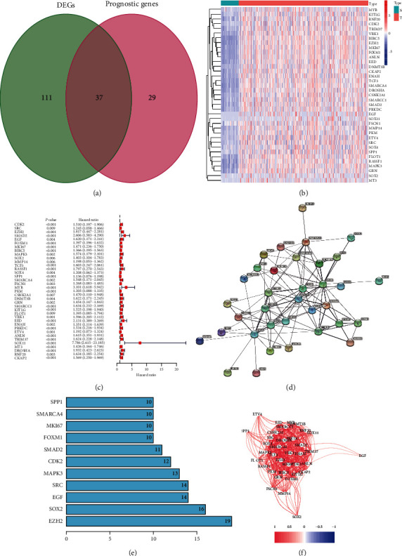
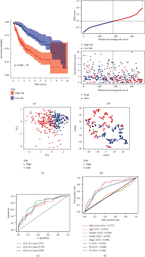
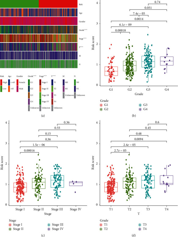
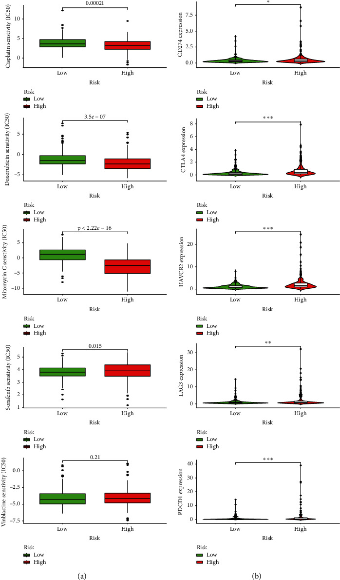
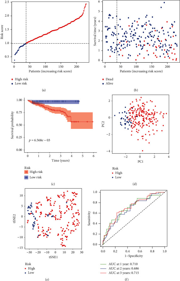
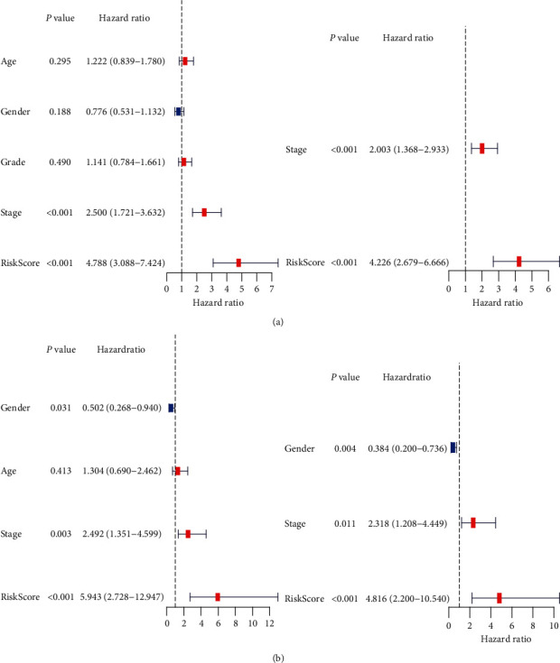
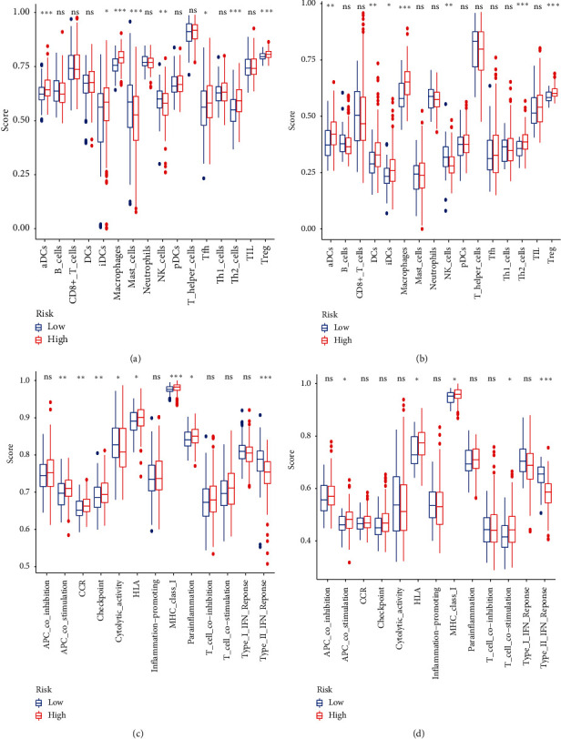
Results: There were 148 differentially expressed MRGs (DEMRGs), out of which 37 MRGs were found associated with overall survival (OS) in the univariate Cox analysis. A novel signature integrating of 5 MRGs was constructed, which split patients into high- and low-risk groups. Kaplan-Meier analysis revealed that high-risk patients had unfavorable OS than those low-risk counterparts. Receiver operating characteristic curve (ROC) showed great performance of this signature in predictive ability. Multivariate Cox analysis confirmed that this signature could independently predict HCC prognosis. The analysis of immune cell infiltration demonstrated that immune status varied differently between high- and low-risk groups. The analysis of clinicopathological characteristics suggested that tumor grade, clinical stage, and T stage were different between risk groups. The analysis between this signature and chemotherapeutic efficacy and immunosuppressive molecules indicated that this signature could serve as a promising predictor.

Conclusions: In conclusion, we constructed and verified a novel signature from the perspective of MET, which was significantly associated with HCC prognosis, clinicopathological features, immune status, chemotherapeutic efficacy, and immunosuppressive biomarkers.


Conflict of interest statement

The authors declare that they have no conflicts of interest.

Figures

Figure 1
Figure 1
Identification of prognosis-related DEMRGs in the TCGA-LIHC dataset. (a) The Venn diagram presenting DEMRGs which were associated with OS in the univariate Cox regression analysis. (b) The heatmap showing 37 prognosis-related DEMRGs. (c) The forest plot displaying the relationship between 37 prognosis-related DEMRGs and OS in the univariate Cox regression analysis. (d) The PPI network among candidate genes obtained from the STRING database. (e) Hub genes with the top 11 degrees of interaction. (f) The correlation analysis of candidate genes.
Figure 2
Figure 2
Development of a MRGs expression signature in the TCGA dataset. (a) The Kaplan–Meier curve survival analysis. (b) The risk score curve plot and risk score scatter plot of high- and low-risk HCC patients. (c) PCA plot of the TCGA dataset. (d) t-SNE analysis of the TCGA dataset. (e) AUC of time-dependent ROC used to assess performance of this signature in predictive ability. (f) AUC of this signature and clinicopathological parameters.
Figure 3
Figure 3
Clinicopathological features of this novel expression signature. Clinical characteristics (a), including tumor grade (b), clinical stage (c), and T stage (d) significantly associated with the risk and risk score.
Figure 4
Figure 4
The relationship between this signature and chemotherapeutic efficacy and ICIs-related molecules. (a) Low-risk group having a higher IC50 of cisplatin, doxorubicin, and mitomycin and a lower IC50 of sorafenib. (b) High-risk group positively related with PDCD1, CD274, CTLA4, HAVCR2, and LAG3.
Figure 5
Figure 5
Validation of this novel expression signature in the ICGC dataset. (a) The risk score curve plot in the ICGC dataset. (b) The risk score scatter plot of high- and low-risk HCC patients. (c) The Kaplan–Meier curve survival analysis. (d) PCA plot of the ICGC dataset. (e) t-SNE analysis of the ICGC dataset. (f) AUC of time-dependent ROC used to assess performance of this signature in predictive ability in the ICGC dataset.
Figure 6
Figure 6
Results of univariate and multivariate Cox regression analysis of OS in the TCGA development dataset (a) and the ICGC validation dataset (b).
Figure 7
Figure 7
Representative results of GO and KEGG analyses. The most significant GO enrichment and KEGG pathways in the TCGA dataset (a, c) and ICGC dataset (b, d) are displayed.
Figure 8
Figure 8
Comparison of the ssGSEA scores between different risk groups in the TCGA dataset and ICGC dataset. The scores of 16 immune cells (a, b) and 13 immune-related functions (c, d) are displayed in boxplots. Adjusted p values are shown. ns, not significant. P < 0.05, ∗∗P < 0.01, ∗∗∗P < 0.001.

Similar articles

Cited by

References

    1. Forner A., Reig M., Bruix J. Hepatocellular carcinoma. The Lancet. 2018;391(10127):1301–1314. doi: 10.1016/s0140-6736(18)30010-2. - DOI - PubMed
    1. Sim H.-W., Knox J. Hepatocellular carcinoma in the era of immunotherapy. Current Problems in Cancer. 2018;42(1):40–48. doi: 10.1016/j.currproblcancer.2017.10.007. - DOI - PubMed
    1. Villanueva A. Hepatocellular carcinoma. New England Journal of Medicine. 2019;380(15):1450–1462. doi: 10.1056/nejmra1713263. - DOI - PubMed
    1. Hartke J., Johnson M., Ghabril M. The diagnosis and treatment of hepatocellular carcinoma. Seminars in Diagnostic Pathology. 2017;34(2):153–159. doi: 10.1053/j.semdp.2016.12.011. - DOI - PubMed
    1. Pei D., Shu X., Gassama-Diagne A., Thiery J. P. Mesenchymal-epithelial transition in development and reprogramming. Nature Cell Biology. 2019;21(1):44–53. doi: 10.1038/s41556-018-0195-z. - DOI - PubMed

LinkOut - more resources