. 2021 Jul 27:2021:9966966.
doi: 10.1155/2021/9966966. eCollection 2021.

Comparison between Intra-Articular Injection of Infrapatellar Fat Pad (IPFP) Cell Concentrates and IPFP-Mesenchymal Stem Cells (MSCs) for Cartilage Defect Repair of the Knee Joint in Rabbits

Affiliations

Comparison between Intra-Articular Injection of Infrapatellar Fat Pad (IPFP) Cell Concentrates and IPFP-Mesenchymal Stem Cells (MSCs) for Cartilage Defect Repair of the Knee Joint in Rabbits

Yaguang Han et al. Stem Cells Int. .

Abstract

Mesenchymal stem cells (MSCs) have emerged as a promising therapeutic method in regenerative medicine. Our previous research adopted a simple nonenzymatic strategy for the preparation of a new type of ready-to-use infrapatellar fat pad (IPFP) cell concentrates. The aim of this study was to compare the therapeutic efficacy of intra-articular (IA) injection of autologous IPFP cell concentrates and allogeneic IPFP-MSCs obtained from these concentrates in a rabbit articular cartilage defect model. IPFP-MSCs sprouting from the IPFP cell concentrates were characterized via flow cytometry as well as based on their potential for differentiation into adipocytes, osteoblasts, and chondrocytes. In the rabbit model, cartilage defects were created on the trochlear groove, followed by treatment with IPFP cell concentrates, IPFP-MSCs, or normal saline IA injection. Distal femur samples were evaluated at 6 and 12 weeks posttreatment via macroscopic observation and histological assessment based on the International Cartilage Repair Society (ICRS) macroscopic scoring system as well as the ICRS visual histological assessment scale. The macroscopic score and histological score were significantly higher in the IPFP-MSC group compared to the IPFP cell concentrate group at 12 weeks. Further, both treatment groups had higher scores compared to the normal saline group. In comparison to the latter, the groups treated with IPFP-MSCs and IPFP cell concentrates showed considerably better cartilage regeneration. Overall, IPFP-MSCs represent an effective therapeutic strategy for stimulating articular cartilage regeneration. Further, due to the simple, cost-effective, nonenzymatic, and safe preparation process, IPFP cell concentrates may represent an effective alternative to stem cell-based therapy in the clinic.


Conflict of interest statement

The authors declare that there is no conflict of interest regarding the publication of this paper.

Figures

Figure 1
Figure 1
The microscopical appearance of the third passage generation of IPFP-MSCs from the (a, b) IPFP-MSC group and the (c, d) cell concentrate group exhibited typical spindle-shaped morphology and cells were anchorage dependent.
Figure 2
Figure 2
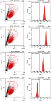
The surface markers of IPFP-MSCs isolated from three rabbits characterized by flow cytometry in passage 3: no expression of CD45 and strong expression of CD44, CD90, and CD105.
Figure 3
Figure 3
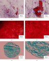
The three-way differentiation and staining results of IPFP-MSCs isolated from three rabbits. (a, b) After staining with Oil red O for adipogenic differentiation, red lipid droplets fused into pieces can be seen. (c, d) After staining with Alizarin red for osteogenic differentiation, there were red calcium nodules fused into masses. (e, f) After staining with Alcian blue for chondrogenic differentiation, a large amount of blue-stained acid mucopolysaccharide appears among the cells.
Figure 4
Figure 4
(a) The gross appearance of cartilage repair in vivo at 6 and 12 weeks after surgery. (b) The ICRS macroscopic evaluation scores of articular cartilage repair. (All data were expressed as the mean ± standard deviation (SD). Comparisons were performed with the Student t-test or one-way ANOVA for experiments with more than two subgroups. Asterisks indicate significant differences (P < 0.05; ∗∗P < 0.01) compared with the corresponding control).
Figure 5
Figure 5
(a, b) Toluidine blue and Safranin O/Fast Green staining of different groups at 6 and 12 weeks. (c) The ICRS visual histological assessment scores of articular cartilage repair. (All data were expressed as the mean ± SD. Comparisons were performed with the Student t-test or one-way ANOVA for experiments with more than two subgroups. Asterisks indicate significant differences (P < 0.05; ∗∗P < 0.01) compared with the corresponding control).

Similar articles

Cited by

References

    1. Lieberthal J., Sambamurthy N., Scanzello C. R. Inflammation in joint injury and post-traumatic osteoarthritis. Osteoarthritis and Cartilage. 2015;23:1825–1834. doi: 10.1016/j.joca.2015.08.015. - DOI - PMC - PubMed
    1. Armiento A. R., Stoddart M. J., Alini M., Eglin D. Biomaterials for articular cartilage tissue engineering: learning from biology. Acta Biomaterialia. 2018;65:1–20. doi: 10.1016/j.actbio.2017.11.021. - DOI - PubMed
    1. Jiang S., Guo W., Tian G., et al. Clinical application status of articular cartilage regeneration techniques: tissue-engineered cartilage brings new hope. Stem Cells International. 2020;2020:16. doi: 10.1155/2020/5690252.5690252 - DOI - PMC - PubMed
    1. Block J. A., Cherny D. Management of knee osteoarthritis: what internists need to know. The Medical Clinics of North America. 2021;105:367–385. doi: 10.1016/j.mcna.2020.10.005. - DOI - PubMed
    1. Katz J. N., Arant K. R., Loeser R. F. Diagnosis and treatment of hip and knee osteoarthritis: a review. JAMA. 2021;325:568–578. doi: 10.1001/jama.2020.22171. - DOI - PMC - PubMed

LinkOut - more resources