. 2018 Nov;13(11):803-818.
doi: 10.2217/fvl-2018-0071. Epub 2018 Oct 24.

Molecular features and translational outlook for Epstein-Barr virus-associated gastric cancer

Affiliations

Molecular features and translational outlook for Epstein-Barr virus-associated gastric cancer

Lyla J Stanland et al. Future Virol. 2018 Nov.

Abstract

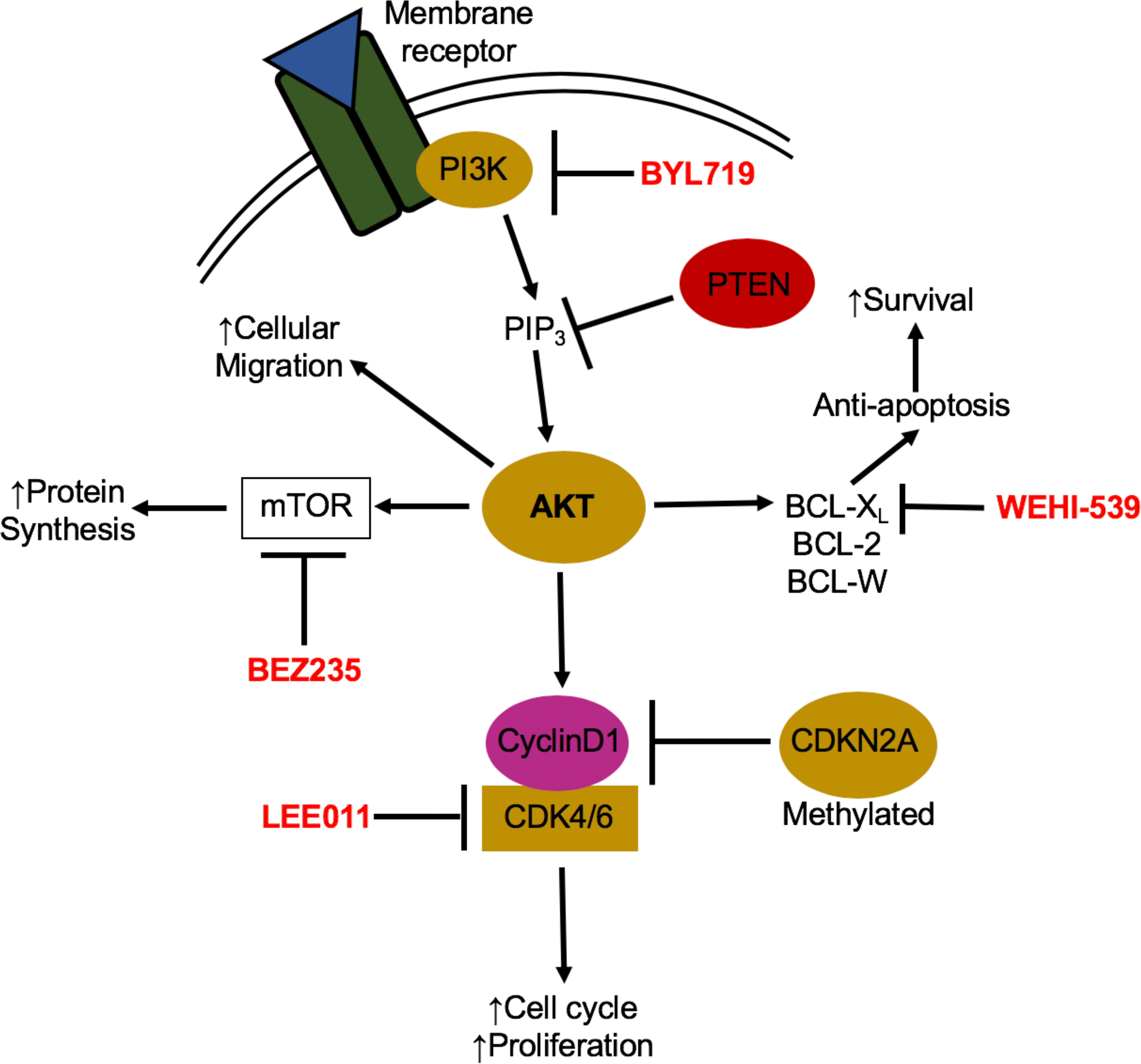
Epstein-Barr Virus (EBV) was the first discovered human tumor virus and is the etiological agent of B cell lymphomas and also epithelial cancers. Indeed, nearly 10% of gastric cancers worldwide are EBV-positive and display unique molecular, epigenetic, and clinicopathological features. EBV-positive gastric cancers display the highest rate of host genome methylation of all tumor types studied and harbor recurrent mutations activating PI3Kα, silencing ARID1A, and amplifying PD-L1. While EBV infection of B cells can be studied efficiently, de novo epithelial cell infection is much more difficult. We propose that new culture models including 3D-based gastric organoids and xenografts can bring new insight into EBV-induced gastric carcinogenesis and will lead to improved precision medicine-based therapies for patients with EBV-positive gastric cancer.

Keywords: EBV; Epstein-Barr virus; PI3K; gastric cancer; immunotherapy; methylation.


Figures

Figure 1:
Figure 1:. Key features of EBV-associated gastric cancer
EBV-positive gastric cancers are molecularly, pathologically and epigenetically distinct from EBV-negative gastric cancers. EBV-associated gastric tumors display an extreme CpG island hypermethylator phenotype (CIMP), and are the most hypermethylated of all tumor types. EBV-positive tumors display an 80% rate of PIK3CA activating mutation and 55% rate of ARID1A silencing mutation which frequently co-occurs with PTEN inactivation. Tumors are made up of EBV-positive gastric epithelial cells that up-regulate PD-L1 and densely infiltrating lymphocytes contributing to an inflammatory microenvironment. EBV infected gastric cells display an extremely restricted latency profile where only a few crucial proteins are expressed.
Figure 2:
Figure 2:. PI3K pathway signaling and drug treatments
Activation of the PI3K/AKT signaling pathway occurs through binding of an external stimuli to a cellular receptor such as a receptor tyrosine kinase, cytokine receptor or G-coupled protein receptor. Mutations in the catalytic domain of PI3K, PIK3CA, can lead to constitutive activation of the pathway resulting in increased proliferation, protein synthesis, and cell survival and migration. When an external stimulus binds to its appropriate cellular receptor, PI3K is recruited to the intracellular domain of the receptor and phosphorylates PIP2 to PIP3. PIP3 then activates AKT leading to a number of downstream protein functions including activation of cyclin D1, mTOR and BCL-xL. There are a number of developed drugs that target different members of the PI3K/AKT pathway and have been shown to be effective in PIK3CA mutant breast cancers and can potentially be used in PIK3CA mutant EBV-positive gastric cancers as well.
Figure 3:
Figure 3:. Mechanism of hypermethylation in EBV-associated gastric cancer
CpG islands located upstream of the coding region are normally non-methylated to allow for transcription, but are methylated by DNA-methyltransferases (DNMTs) to control gene transcription and expression. EBV infection of gastric epithelial cells induces hypermethylation by taking advantage of existing cellular methylation mechanisms. A) Cytosine bases are methylated on the 5-position carbon by DNMTs, changing 5-C to 5mC. Methylated cytosines can be oxidized by ten-eleven translocation (TET) family enzymes changing 5mC to 5hmC. B) CpG islands are located throughout promoter regions and surrounding the transcription start sites (TSS) of genes. CpG islands are normally un-methylated to allow for transcription of genes including tumor suppressors such as PTEN and cell cycle genes including CDKN2A. C) Virally encoded LMP2A can phosphorylate transcription factor STAT3, which moves to the nucleus and activates transcription of DNMT1 leading to induction of hypermethylation. Additionally, LMP2A inhibits TET2 oxidation of methylated cytosines to maintain hypermethylation phenotype.
Figure 4:
Figure 4:. Culture model systems in which to study tumorigenesis and potential therapies for EBVaGC
EBV does not readily infect and establish latency in epithelial cells in vitro and multiple culture model systems are in development to allow for study of EBVaGC tumorigenesis both in vitro and in vivo. A) Naturally occurring EBV-positive GaCa-derived cell lines can be treated with PI3K pathway drugs shown to be effective in other solid tumors and cellular growth is assayed via growth inhibition assays and xenograft tumor volume and growth. B) Using the co-culture method, EBV negative GaCa cell lines can be infected with rEBV and selected for using drug resistance and GFP expression. In vitro infected cells can also be treated with PI3K pathway drugs to assay growth and cell death. Additionally, EBV infected cell lines can be sequenced to understand induction of hypermethylation and genomic and transcriptomic changes that occur during infection. C) Stem cells can be differentiated into gastric organoids or directly infected to study proliferation and survival of differentiated cell types of the stomach.
Figure 5:
Figure 5:. PD-1 blockade therapy for EBV-associated gastric tumors
Disruption of PD-1/PD-L1 interactions between gastric tumor cells and infiltrating CD8+ T cells can be an effective therapy for the treatment of EBV-positive gastric cancers due to overexpression of PD-L1 and a IFNγ dominated inflammatory microenvironment. A) PD-L1 on tumor cells is recognized by PD-1 expressed by cytotoxic T cells preventing normal T cell effector functions even if the TCR is presented with a tumor specific antigen. Through this interaction, the tumor cells can evade the immune response. B) Anti-PD-1 antibodies can be used to prevent binding of PD-L1 to PD-1 allowing the CD8 T cells to recognize tumor cells through antigen presentation and perform effector functions to kill tumor cells.

References

    1. Young LS, Yap LF & Murray PG. Epstein-Barr virus: more than 50 years old and still providing surprises. Nat. Rev. Cancer 16, 789–802, (2016). - PubMed
    1. Niedobitek G, Agathanggelou A, Steven N & Young LS. Epstein-Barr virus (EBV) in infectious mononucleosis: detection of the virus in tonsillar B lymphocytes but not in desquamated oropharyngeal epithelial cells. Mol. Pathol 53, 37–42, (2000). - PMC - PubMed
    1. Price AM et al. Epstein-Barr virus ensures B cell survival by uniquely modulating apoptosis at early and late times after infection. Elife 6, (2017). - PMC - PubMed
    1. Young LS & Dawson CW. Epstein-Barr virus and nasopharyngeal carcinoma. Chin. J. Cancer 33, 581–590, (2014). - PMC - PubMed
    1. Epstein M, Achong B & Barr Y Virus particles in cultured lymphoblasts from Burkitt’s lymphoma. Lancet 1, (1964). - PubMed

LinkOut - more resources