

COVID-19 and pulmonary tuberculosis - A diagnostic dilemma
- PMID: 34367387
- PMCID: PMC8326013
- DOI: 10.1016/j.radcr.2021.07.079
COVID-19 and pulmonary tuberculosis - A diagnostic dilemma
Abstract
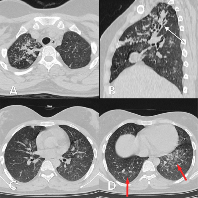
Coronavirus disease 2019 (COVID-19) is an infectious disease caused by the severe acute respiratory syndrome coronavirus 2 (SARS-CoV-2). Meanwhile, pulmonary tuberculosis(TB) is one of the most common infective lung diseases in developing nations. The concurrence of pulmonary TB and COVID-19 can lead to poor prognosis, owing to the pre-existing lung damage caused by TB. Case presentation: We describe the imaging findings in 3 cases of COVID-19 pneumonia with co-existing pulmonary TB on HRCT thorax. The concurrence of COVID-19 and pulmonary TB can be a diagnostic dilemma. Correct diagnosis and prompt management is imperative to reduce mortality and morbidity. Hence it is pertinent for imaging departments to identify and report these distinct entities when presenting in conjunction.
Keywords: AFB, Acid-fast bacilli; CO-RADS, COVID-19 Reporting and Data System; COVID -19; COVID-19, Coronavirus disease 2019; CRP, C-reactive protein; CT, Computed tomography; Case report; Co-infection; DNA, Deoxyribonucleic acid; DOTS, Directly Observed Therapy, Short-Course; GGOs, Ground glass opacities; Ground glass opacities; HRCT; HRCT, High resolution computed tomography; ICU, Intensive care unit; RT-PCR, Reverse transcriptase-polymerase chain reaction; SARS-CoV-2, Severe acute respiratory syndrome coronavirus 2; TB, Tuberculosis; Tuberculosis; WBC, White blood cell.
© 2021 The Authors. Published by Elsevier Inc. on behalf of University of Washington.
Figures


Similar articles
-
CO RADS grade of HRCT Thorax and RT PCR testing for the diagnosis of coronavirus disease 2019 (COVID 19): A descriptive hospital based study of asymptomatic planned surgery cases.Ann Afr Med. 2023 Jan-Mar;22(1):40-44. doi: 10.4103/aam.aam_205_21. Ann Afr Med. 2023. PMID: 36695220 Free PMC article.
-
Clinical, laboratory and high-resolution computed tomography (HRCT) thorax profile of reverse transcription polymerase chain reaction (RT-PCR) negative COVID-19 suspects with moderate to severe disease.J Educ Health Promot. 2022 Oct 31;11:333. doi: 10.4103/jehp.jehp_287_22. eCollection 2022. J Educ Health Promot. 2022. PMID: 36568002 Free PMC article.
-
A 28-Year-Old Man from India with SARS-Cov-2 and Pulmonary Tuberculosis Co-Infection with Central Nervous System Involvement.Am J Case Rep. 2020 Aug 19;21:e926034. doi: 10.12659/AJCR.926034. Am J Case Rep. 2020. PMID: 32813683 Free PMC article.
-
Tuberculosis and COVID-19: A combined global threat to human civilization.Clin Epidemiol Glob Health. 2022 May-Jun;15:101031. doi: 10.1016/j.cegh.2022.101031. Epub 2022 Mar 27. Clin Epidemiol Glob Health. 2022. PMID: 35372717 Free PMC article. Review.
-
Disruption of CCR5 signaling to treat COVID-19-associated cytokine storm: Case series of four critically ill patients treated with leronlimab.J Transl Autoimmun. 2021;4:100083. doi: 10.1016/j.jtauto.2021.100083. Epub 2021 Jan 6. J Transl Autoimmun. 2021. PMID: 33521616 Free PMC article. Review.
Cited by
-
The Impact of COVID-19 on the Tuberculosis Features in a Romanian Pneumology Hospital.J Multidiscip Healthc. 2024 May 22;17:2489-2498. doi: 10.2147/JMDH.S463859. eCollection 2024. J Multidiscip Healthc. 2024. PMID: 38799014 Free PMC article.
-
The COVID-19/Tuberculosis Syndemic and Potential Antibody Therapy for TB Based on the Lessons Learnt From the Pandemic.Front Immunol. 2022 Feb 15;13:833715. doi: 10.3389/fimmu.2022.833715. eCollection 2022. Front Immunol. 2022. PMID: 35242137 Free PMC article. Review.
-
COVID-19 and Tuberculosis: Unveiling the Dual Threat and Shared Solutions Perspective.J Clin Med. 2023 Jul 19;12(14):4784. doi: 10.3390/jcm12144784. J Clin Med. 2023. PMID: 37510899 Free PMC article.
-
Impact of Severity of COVID-19 in TB Disease Patients: Experience from an Italian Infectious Disease Referral Hospital.Infect Dis Rep. 2025 Feb 5;17(1):11. doi: 10.3390/idr17010011. Infect Dis Rep. 2025. PMID: 39997463 Free PMC article.
-
Effect of Concomitant Tuberculosis Infection on COVID-19 Disease in Children: A Matched, Retrospective Cohort Study.J Trop Pediatr. 2022 Jun 6;68(4):fmac056. doi: 10.1093/tropej/fmac056. J Trop Pediatr. 2022. PMID: 35796754 Free PMC article.
References
-
- World Health Organization. Coronavirus. https://www.who.int/health-topics/coronavirus#tab=tab_1.Date of access June 20, 2021.
-
- Lu R, Zhao X, Li J, Niu P, Yang B, Wu H. Genomic characterisation and epidemiology of 2019 novel coronavirus: implications for virus origins and receptor binding. Lancet. 2020;395(10224):565–574. doi: 10.1016/S0140-6736(20)30251-8. Epub 2020 Jan 30. PMID: 32007145; PMCID: PMC7159086. - DOI - PMC - PubMed
Publication types
LinkOut - more resources
Full Text Sources
Research Materials
Miscellaneous
