

JOINT SEGMENTATION OF MULTIPLE SCLEROSIS LESIONS AND BRAIN ANATOMY IN MRI SCANS OF ANY CONTRAST AND RESOLUTION WITH CNNs
- PMID: 34367472
- PMCID: PMC8340983
- DOI: 10.1109/isbi48211.2021.9434127
JOINT SEGMENTATION OF MULTIPLE SCLEROSIS LESIONS AND BRAIN ANATOMY IN MRI SCANS OF ANY CONTRAST AND RESOLUTION WITH CNNs
Abstract
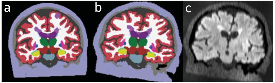
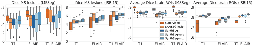
We present the first deep learning method to segment Multiple Sclerosis lesions and brain structures from MRI scans of any (possibly multimodal) contrast and resolution. Our method only requires segmentations to be trained (no images), as it leverages the generative model of Bayesian segmentation to generate synthetic scans with simulated lesions, which are then used to train a CNN. Our method can be retrained to segment at any resolution by adjusting the amount of synthesised partial volume. By construction, the synthetic scans are perfectly aligned with their labels, which enables training with noisy labels obtained with automatic methods. The training data are generated on the fly, and aggressive augmentation (including artefacts) is applied for improved generalisation. We demonstrate our method on two public datasets, comparing it with a state-of-the-art Bayesian approach implemented in FreeSurfer, and dataset specific CNNs trained on real data. The code is available at https://github.com/BBillot/SynthSeg.
Keywords: MS lesion; contrast-agnostic; segmentation.
Figures

References
-
- Confavreux C et al. “Rate of Pregnancy-Related Relapse in MS,” J. of Med, vol. 339, pp. 285–291, 1998. - PubMed
-
- Fisher E et al. “Gray matter atrophy in multiple sclerosis study,” Annals of Neur, vol. 64, pp. 255–265, 2008. - PubMed
-
- Barkhof F et al. “Imaging outcomes for neuroprotection and repair in multiple sclerosis trials,” Nature Reviews. Neurology, 5, pp. 256–266, 2009. - PubMed
-
- Kamnitsas K et al. “Efficient multi-scale 3D CNN with fully connected CRF for accurate brain lesion segmentation,” Medical Im. Analysis, vol. 36, pp. 61–78, 2017. - PubMed
Grants and funding
LinkOut - more resources
Full Text Sources
Research Materials