Case Reports
. 2021 Jul 26;13(7):944-970.
doi: 10.4252/wjsc.v13.i7.944.

First immunohistochemical evidence of human tendon repair following stem cell injection: A case report and review of literature

Affiliations
Case Reports

First immunohistochemical evidence of human tendon repair following stem cell injection: A case report and review of literature

Eckhard Alt et al. World J Stem Cells. .

Abstract

Background: Current clinical treatment options for symptomatic, partial-thickness rotator cuff tear (sPTRCT) offer only limited potential for true tissue healing and improvement of clinical results. In animal models, injections of adult stem cells isolated from adipose tissue into tendon injuries evidenced histological regeneration of tendon tissue. However, it is unclear whether such beneficial effects could also be observed in a human tendon treated with fresh, uncultured, autologous, adipose derived regenerative cells (UA-ADRCs). A specific challenge in this regard is that UA-ADRCs cannot be labeled and, thus, not unequivocally identified in the host tissue. Therefore, histological regeneration of injured human tendons after injection of UA-ADRCs must be assessed using comprehensive, immunohistochemical and microscopic analysis of biopsies taken from the treated tendon a few weeks after injection of UA-ADRCs.

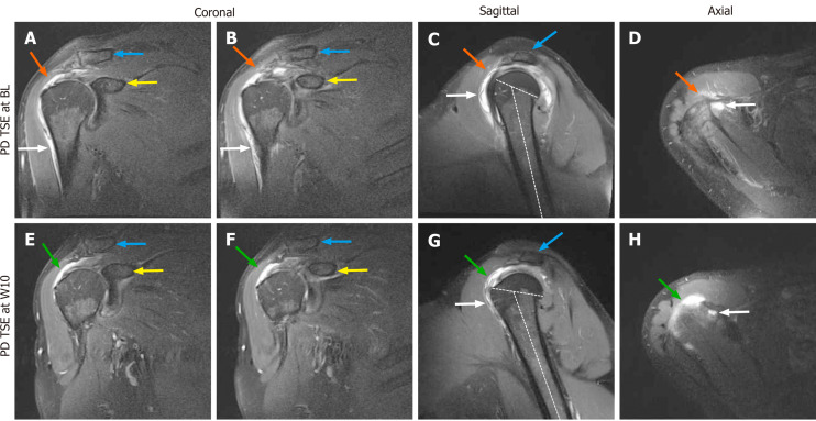
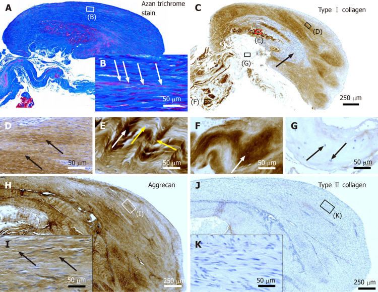
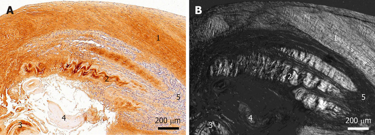
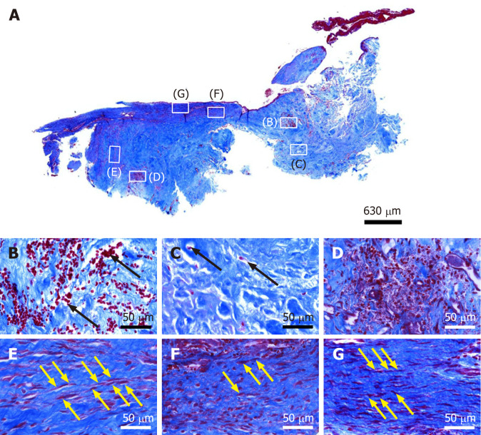
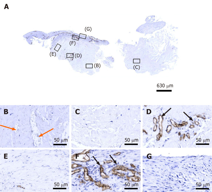
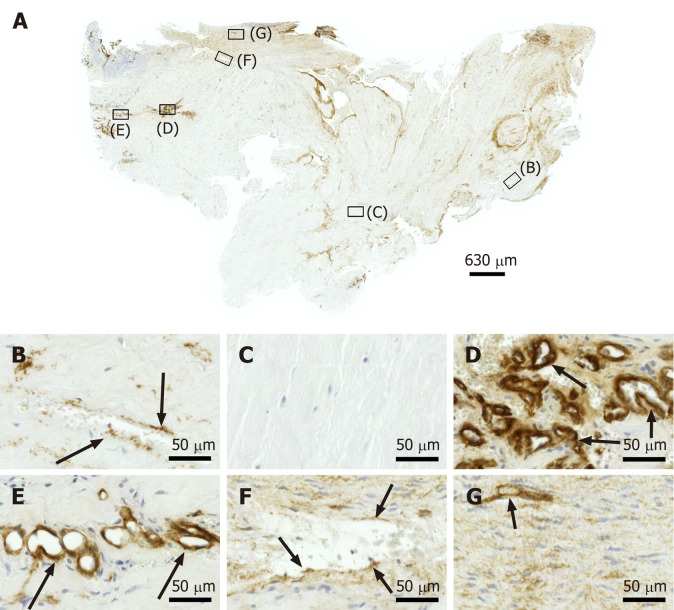
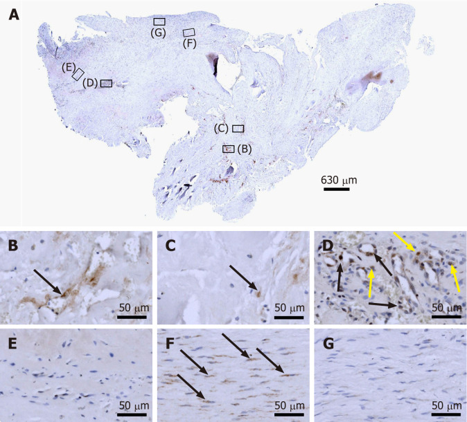
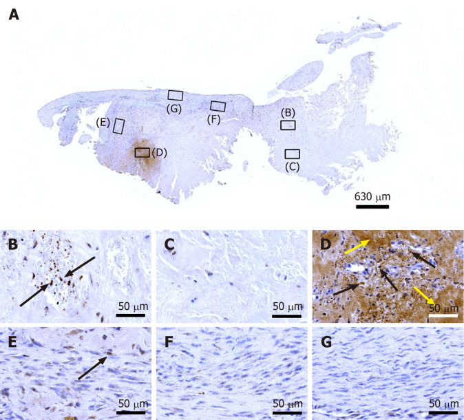
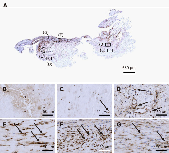
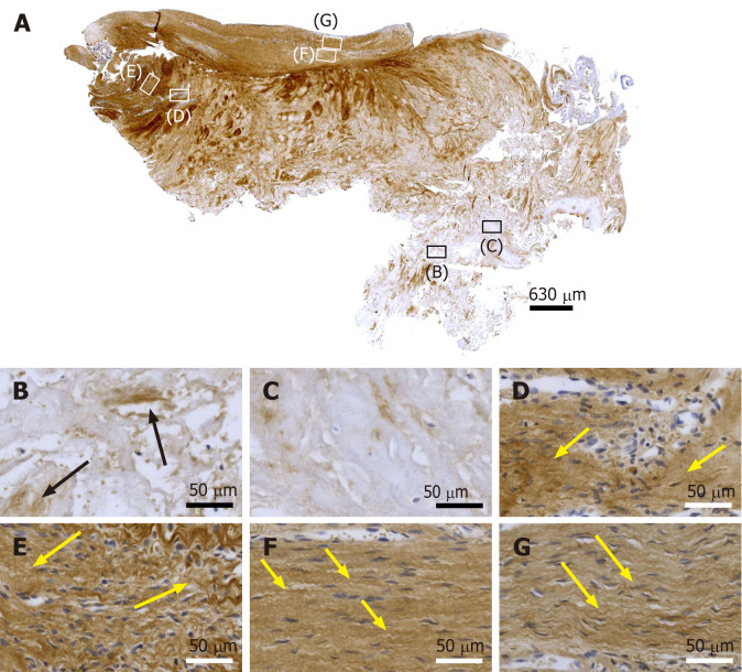
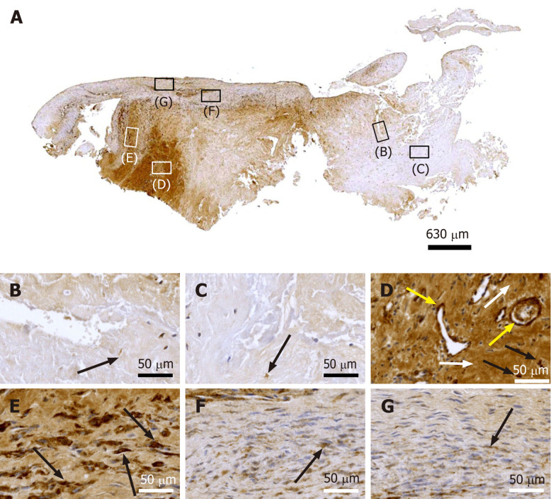
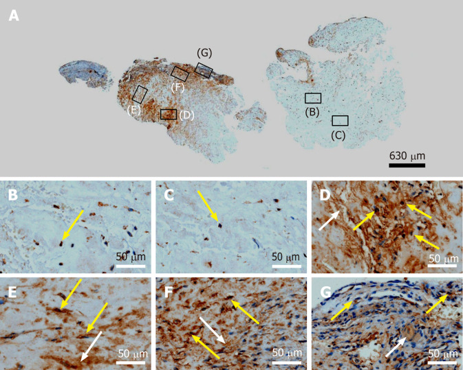
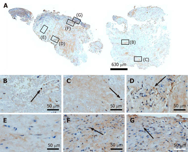
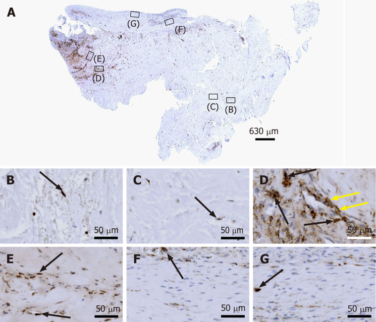
Case summary: A 66-year-old patient suffered from sPTRCT affecting the right supraspinatus and infraspinatus tendon, caused by a bicycle accident. On day 18 post injury [day 16 post magnetic resonance imaging (MRI) examination] approximately 100 g of abdominal adipose tissue was harvested by liposuction, from which approximately 75 × 106 UA-ADRCs were isolated within 2 h. Then, UA-ADRCs were injected (controlled by biplanar X-ray imaging) adjacent to the injured supraspinatus tendon immediately after isolation. Despite fast clinical recovery, a follow-up MRI examination 2.5 mo post treatment indicated the need for open revision of the injured infraspinatus tendon, which had not been treated with UA-ADRCs. During this operation, a biopsy was taken from the supraspinatus tendon at the position of the injury. A comprehensive, immunohistochemical and microscopic analysis of the biopsy (comprising 13 antibodies) was indicative of newly formed tendon tissue.

Conclusion: Injection of UA-ADRCs can result in regeneration of injured human tendons by formation of new tendon tissue.

Keywords: Adipose derived regenerative cells; Case report; Cell-based therapy at point of care; Partial-thickness rotator cuff tear; Stem cells; Tendon regeneration.


Conflict of interest statement

Conflict-of-interest statement: Alt EU is Executive Chair of InGeneron, Inc. (Houston, TX) and Chairman of the Board of Isar Klinikum (Munich, Germany). Alt C is Director of Science and Research of InGeneron GmbH (Munich, Germany) and of SciCoTec (Grünwald, Germany), the principal shareholder of InGeneron, Inc., which owns InGeneron GmbH (Munich, Germany). Schmitz C served as consultant to SciCoTec and the Alliance of Cardiovascular Researchers, and is Advisory Medical Director of InGeneron, Inc.

Figures

Figure 1
Figure 1
Proton density weighted, fat saturated, Turbo Spin Echo, 1.5 T magnetic resonance imaging scans of the right shoulder of the investigated patient (matrix 320 × 320; slice thickness 3 mm; inter-slice gap 0.3 mm; echo time 37 ms; repetition time 2450 ms at baseline and 3480 ms at 10 wk post injection). A and B: Coronal scans at baseline (BL) (white arrows, trauma-related bruising; orange arrows, position of the supraspinatus tendon at which a hyperintense structure was found at 10 wk post injection (W10) but not at BL; blue arrows, clavicle; yellow arrows, coracoid process of the scapula); C: Sagittal scan at BL (white arrow, trauma-related bruising; orange arrow, position of the supraspinatus tendon at which a hyperintense structure was found at W10 but not at BL; blue arrow, acromion; dotted lines, long axis of the humeral shaft and delineation of the humeral head, with same angle between the lines as in Panel G); D: Axial scan at BL (white arrow, trauma-related bruising; orange arrow, position of the supraspinatus tendon at which a hyperintense structure was found at W10 but not at BL); E and F: Coronal scans at W10 (green arrows, hyperintense structure at the position of the supraspinatus tendon that was found at W10 but not at BL; blue arrows, clavicle; yellow arrows, coracoid process of the scapula); G: Sagittal scan at W10 (white arrow, trauma-related bruising; green arrow, hyperintense structure at the position of the supraspinatus tendon that was found at W10 but not at BL; blue arrow, acromion; dotted lines, long axis of the humerus and delineation of the humeral head, with same angle between the lines as in Panel C); H: Axial scan at W10 (white arrow, trauma-related bruising; green arrow, hyperintense structure at the position of the supraspinatus tendon that was found at W10 but not at BL). BL: Baseline; PD TSE: Proton density weighted turbo spin echo; W10: 10 wk post injection.
Figure 2
Figure 2
Histological and immunohistochemical analysis of representative sections from the first part of the biopsy that was investigated in this study. A and B: Section stained with Azan trichrome stain; cells are in red and collagen is in blue (inset in Panel A, position of the high-power photomicrograph displayed in Panel B; white arrows in Panel B, elongated, fibroblast-like cells (tenocytes) arranged in long and parallel chains between collagen fibers); C-G: Immunohistochemical detection of type I collagen in a section adjacent to the one shown in Panel A; counterstaining was performed with Mayer's hematoxylin (black arrow in Panel C, region with almost complete absence of immunolabeling for type I collagen but a high cell density; insets in Panel C, position of the high-power photomicrographs displayed in Panels D-G, showing the following four different regions: D: Immunolabeling for organized, slightly undulating type I collagen (black arrows in Panel D) and high cell density; E: Immunolabeling for organized type I collagen with discernible crimp arrangement (white arrow in Panel E) and a few cells (yellow arrows in Panel E); F: Immunolabeling for organized type I collagen with discernible crimp arrangement (white arrow in Panel F) and absence of cells; G: Absence of immunolabeling for type I collagen and a few, rounded cells (black arrows in Panel G); H and I: Immunohistochemical detection of aggrecan in a section adjacent to the ones shown in Panels A and C of the same biopsy; counterstaining was performed with Mayer's hematoxylin (inset in Panel H, position of the high-power photomicrograph displayed in Panel I; black arrows in Panel I, immunolabeling for aggrecan); J and K: Immunohistochemical detection of type II collagen in another section adjacent to the ones shown in Panels A, C and H of the same biopsy; counterstaining was also performed with Mayer's hematoxylin (inset in Panel J, position of the high-power photomicrograph displayed in Panel K).
Figure 3
Figure 3
Histological and immunohistochemical analysis of a representative section from the first part of the biopsy that was investigated in this study. A: Immunohistochemical detection of type I collagen, showing the following 5 different regions (high-power photomicrographs are provided in Figure 2): 1Organized, slightly undulating type I collagen and high cell density; 2Organized type I collagen with discernible crimp arrangement and a few cells; 3Organized type I collagen with discernible crimp arrangement and almost complete absence of cells; 4Almost complete absence of immunolabeling for type I collagen and a few, rounded cells; and 5Almost complete absence of immunolabeling for type I collagen but a high cell density; B: Corresponding polarized light microscopic image of the same field of view. Note the clear difference in collagen fiber birefringence between regions 1 and 2, and the absence of collagen fiber birefringence in regions 4 and 5.
Figure 4
Figure 4
Histological analysis of a representative section of the second part of the biopsy that was investigated in this study (section stained with Azan trichrome stain; cells are in red and collagen is in blue). A: Low-power overview (insets, position of the high-power photomicrographs displayed in Panels B-G); B: Degenerative tendon tissue with formation of microvessels (black arrows, blood cells); C: Degenerative tendon tissue without formation of microvessels (black arrows, rounded cells); D: Spot with very high density of cells and microvessels; E: Tendon tissue in the depth of the biopsy (yellow arrows, elongated, fibroblast-like cells (tenocytes) arranged in long and parallel chains between collagen fibers); F: Tendon tissue below an outer surface of the biopsy (yellow arrows, elongated, fibroblast-like cells (tenocytes) arranged in long and parallel chains between collagen fibers); G: Tendon tissue at an outer surface of the biopsy (yellow arrows, elongated, fibroblast-like cells (tenocytes) arranged in long and parallel chains between collagen fibers).
Figure 5
Figure 5
Immunohistochemical detection of cluster of differentiation 34 in a section of the second part of the biopsy that was investigated in this study (section adjacent to the one shown in Figure 4; counterstaining was performed with Mayer's hematoxylin). A: Low-power overview (insets, position of the high-power photomicrographs displayed in Panels B-G); B: Position of degenerative tendon tissue with formation of microvessels (orange arrows, cells inside a microvessel); C: Position of degenerative tendon tissue without formation of microvessels; D: Position of a spot with very high density of cells and microvessels (black arrows, immunolabeling for cluster of differentiation (CD) 34 in endothelial cells of microvessels); E: Position of tendon tissue in the depth of the biopsy; F: Position of tendon tissue below an outer surface of the biopsy (black arrows, immunolabeling for CD34 in endothelial cells of microvessels); G: Position of tendon tissue at an outer surface of the biopsy.
Figure 6
Figure 6
Immunohistochemical detection of type IV collagen in a section of the second part of the biopsy that was investigated in this study (section adjacent to the one shown in Figure 4; counterstaining was performed with Mayer's hematoxylin). A: Low-power overview (insets, position of the high-power photomicrographs displayed in Panels B-G); B: Position of degenerative tendon tissue with formation of microvessels (black arrows, immunolabeling for type IV collagen in the basement membrane of microvessels); C: position of degenerative tendon tissue without formation of microvessels; D: Position of a spot with very high density of cells and microvessels (black arrows, immunolabeling for type IV collagen in the basement membrane of microvessels); E: Position of tendon tissue in the depth of the biopsy (black arrows, immunolabeling for type IV collagen in the basement membrane of microvessels); F: Position of tendon tissue below an outer surface of the biopsy (black arrows, immunolabeling for type IV collagen in the basement membrane of microvessels); G: Position of tendon tissue at an outer surface of the biopsy (black arrow, immunolabeling for type IV collagen in the basement membrane of microvessels).
Figure 7
Figure 7
Immunohistochemical detection of Ki-67 in a section of the second part of the biopsy that was investigated in this study (section adjacent to the one shown in Figure 4; counterstaining was performed with Mayer's hematoxylin). A: Low-power overview (insets, position of the high-power photomicrographs displayed in Panels B-G); B: Position of degenerative tendon tissue with formation of microvessels (black arrow, intracellular immunolabeling for Ki-67); C: Position of degenerative tendon tissue without formation of microvessels (black arrow, intracellular immunolabeling for Ki-67); D: Position of a spot with very high density of cells and microvessels (black arrows, intracellular immunolabeling for Ki-67 inside microvessel walls; yellow arrows, intracellular immunolabeling for Ki-67 outside microvessel walls); E: Position of tendon tissue in the depth of the biopsy; F: Position of tendon tissue below an outer surface of the biopsy (black arrows, intracellular immunolabeling for Ki-67 in elongated cells in a chain-like arrangement); G: Position of tendon tissue at an outer surface of the biopsy.
Figure 8
Figure 8
Immunohistochemical detection of tenomodulin in a section of the second part of the biopsy that was investigated in this study (section adjacent to the one shown in Figure 4; counterstaining was performed with Mayer's hematoxylin). A: Low-power overview (insets, position of the high-power photomicrographs displayed in Panels B-G); B: Position of degenerative tendon tissue with formation of microvessels (black arrows, intracellular immunolabeling for tenomodulin in cells inside microvessels); C: Position of degenerative tendon tissue without formation of microvessels; D: Position of a spot with very high density of cells and microvessels (black arrows, intracellular immunolabeling for tenomodulin; yellow arrows, extracellular immunolabeling for tenomodulin); E: Position of tendon tissue in the depth of the biopsy (black arrows, intracellular immunolabeling for tenomodulin); F: Position of tendon tissue below an outer surface of the biopsy; G: Position of tendon tissue at an outer surface of the biopsy.
Figure 9
Figure 9
Immunohistochemical detection of type I procollagen in a section of the second part of the biopsy that was investigated in this study (section adjacent to the one shown in Figure 4; counterstaining was performed with Mayer's hematoxylin). A: Low-power overview (insets, position of the high-power photomicrographs displayed in Panels B-G); B: Position of degenerative tendon tissue with formation of microvessels; C: Position of degenerative tendon tissue without formation of microvessels (black arrow, intracellular immunolabeling for type I procollagen); D: Position of a spot with very high density of cells and microvessels (black arrows, intracellular immunolabeling for type I procollagen outside microvessels); E: Position of tendon tissue in the depth of the biopsy (black arrows, intracellular immunolabeling for type I procollagen); F: Position of tendon tissue below an outer surface of the biopsy (black arrows, intracellular immunolabeling for type I procollagen); G: Position of tendon tissue at an outer surface of the biopsy (black arrows, intracellular immunolabeling for type I procollagen).
Figure 10
Figure 10
Immunohistochemical detection of type I collagen in a section of the second part of the biopsy that was investigated in this study (section adjacent to the one shown in Figure 4; counterstaining was performed with Mayer's hematoxylin). A: Low-power overview (insets, position of the high-power photomicrographs displayed in Panels B-G); B: Position of degenerative tendon tissue with formation of microvessels (black arrows, immunolabeling for unorganized type I collagen); C: Position of degenerative tendon tissue without formation of microvessels; D: Position of a spot with very high density of cells and microvessels (yellow arrows, immunolabeling for unorganized type I collagen); E: Position of tendon tissue in the depth of the biopsy (yellow arrows, immunolabeling for unorganized type I collagen); F: Position of tendon tissue below an outer surface of the biopsy (yellow arrows, immunolabeling for organized, slightly undulating type I collagen); G: Position of tendon tissue at an outer surface of the biopsy (yellow arrows, immunolabeling for organized, slightly undulating type I collagen).
Figure 11
Figure 11
Immunohistochemical detection of type III collagen in a section of the second part of the biopsy that was investigated in this study (section adjacent to the one shown in Figure 4; counterstaining was performed with Mayer's hematoxylin). A: Low-power overview (insets, position of the high-power photomicrographs displayed in Panels B-G); B: Position of degenerative tendon tissue with formation of microvessels (black arrows, extracellular immunolabeling for type III collagen); C: Position of degenerative tendon tissue without formation of microvessels; D: Position of a spot with very high density of cells and microvessels (black arrows, extracellular immunolabeling for type III collagen); E: Position of tendon tissue in the depth of the biopsy (black arrows, extracellular immunolabeling for type III collagen); F: Position of tendon tissue below an outer surface of the biopsy; G: Position of tendon tissue at an outer surface of the biopsy.
Figure 12
Figure 12
Immunohistochemical detection of laminin in a section of the second part of the biopsy that was investigated in this study (section adjacent to the one shown in Figure 4; counterstaining was performed with Mayer's hematoxylin). A: Low-power overview (insets, position of the high-power photomicrographs displayed in Panels B-G); B: Position of degenerative tendon tissue with formation of microvessels (black arrow, intracellular immunolabeling for laminin); C: Position of degenerative tendon tissue without formation of microvessels (black arrow, intracellular immunolabeling for laminin); D: Position of a spot with very high density of cells and microvessels (black arrows, intracellular immunolabeling for laminin outside microvessel walls; yellow arrows, intracellular immunolabeling for laminin inside microvessel walls; white arrows, extracellular immunolabeling for laminin); E: Position of tendon tissue in the depth of the biopsy (black arrows, intracellular immunolabeling for laminin); F: Position of tendon tissue below an outer surface of the biopsy (black arrow, intracellular immunolabeling for laminin); G: Position of tendon tissue at an outer surface of the biopsy (black arrow, intracellular immunolabeling for laminin).
Figure 13
Figure 13
Immunohistochemical detection of matrix metalloproteinase 2 (MMP-2) in a section of the second part of the biopsy that was investigated in this study (section adjacent to the one shown in Figure 4; counterstaining was performed with Mayer's hematoxylin). A: Low-power overview (insets, position of the high-power photomicrographs displayed in Panels B-G); B: Position of degenerative tendon tissue with formation of microvessels [yellow arrow, intracellular immunolabeling for matrix metalloproteinase 2 (MMP-2)]; C: Position of degenerative tendon tissue without formation of microvessels (yellow arrow, intracellular immunolabeling for MMP-2); D: Position of a spot with very high density of cells and microvessels (yellow arrows, intracellular immunolabeling for MMP-2; white arrow, extracellular immunolabeling for MMP-2); E: Position of tendon tissue in the depth of the biopsy (yellow arrows, intracellular immunolabeling for MMP-2; white arrow, extracellular immunolabeling for MMP-2); F: Position of tendon tissue below an outer surface of the biopsy (yellow arrows, intracellular immunolabeling for MMP-2; white arrow, extracellular immunolabeling for MMP-2); G: Position of tendon tissue at an outer surface of the biopsy (yellow arrows, intracellular immunolabeling for MMP-2; white arrow, extracellular immunolabeling for MMP-2).
Figure 14
Figure 14
Immunohistochemical detection of matrix metalloproteinase 9 in a section of the second part of the biopsy that was investigated in this study (section adjacent to the one shown in Figure 4; counterstaining was performed with Mayer's hematoxylin). A: Low-power overview (insets, position of the high-power photomicrographs displayed in Panels B-G); B: Position of degenerative tendon tissue with formation of microvessels [black arrow, intracellular immunolabeling for matrix metalloproteinase 9 (MMP-9)]; C: Position of degenerative tendon tissue without formation of microvessels (black arrow, intracellular immunolabeling for MMP-9); D: Position of a spot with very high density of cells and microvessels (black arrow, intracellular immunolabeling for MMP-9); E: Position of tendon tissue in the depth of the biopsy; F: Position of tendon tissue below an outer surface of the biopsy (black arrow, intracellular immunolabeling for MMP-9); G: Position of tendon tissue at an outer surface of the biopsy (black arrow, intracellular immunolabeling for MMP-9).
Figure 15
Figure 15
Immunohistochemical detection of cluster of differentiation 68 in a section of the second part of the biopsy that was investigated in this study (section adjacent to the one shown in Figure 4; counterstaining was performed with Mayer's hematoxylin). A: Low-power overview (insets, position of the high-power photomicrographs displayed in Panels B-G); B: Position of degenerative tendon tissue with formation of microvessels [black arrow, intracellular labeling for cluster of differentiation (CD68)]; C: Position of degenerative tendon tissue without formation of microvessels (black arrow, intracellular labeling for CD68); D: Position of a spot with very high density of cells and microvessels (black arrows, intracellular labeling for CD68 outside microvessel walls; yellow arrows, intracellular immunolabeling for CD68 inside microvessel walls); E: Position of tendon tissue in the depth of the biopsy (black arrows, intracellular labeling for CD68); F: Position of tendon tissue below an outer surface of the biopsy (black arrow, intracellular labeling for CD68); G: Position of tendon tissue at an outer surface of the biopsy (black arrow, intracellular labeling for CD68).

Similar articles

Cited by

References

    1. Matthewson G, Beach CJ, Nelson AA, Woodmass JM, Ono Y, Boorman RS, Lo IK, Thornton GM. Partial Thickness Rotator Cuff Tears: Current Concepts. Adv Orthop. 2015;2015:458786. - PMC - PubMed
    1. Coombes BK, Bisset L, Vicenzino B. Efficacy and safety of corticosteroid injections and other injections for management of tendinopathy: a systematic review of randomised controlled trials. Lancet. 2010;376:1751–1767. - PubMed
    1. Ramírez J, Pomés I, Cabrera S, Pomés J, Sanmartí R, Cañete JD. Incidence of full-thickness rotator cuff tear after subacromial corticosteroid injection: a 12-week prospective study. Mod Rheumatol. 2014;24:667–670. - PubMed
    1. Hurley ET, Hannon CP, Pauzenberger L, Fat DL, Moran CJ, Mullett H. Nonoperative Treatment of Rotator Cuff Disease With Platelet-Rich Plasma: A Systematic Review of Randomized Controlled Trials. Arthroscopy. 2019;35:1584–1591. - PubMed
    1. Schwitzguebel AJ, Kolo FC, Tirefort J, Kourhani A, Nowak A, Gremeaux V, Saffarini M, Lädermann A. Efficacy of Platelet-Rich Plasma for the Treatment of Interstitial Supraspinatus Tears: A Double-Blinded, Randomized Controlled Trial. Am J Sports Med. 2019;47:1885–1892. - PubMed

Publication types