Case Reports
. 2021 Jul 27:2021:8876584.
doi: 10.1155/2021/8876584. eCollection 2021.

Osteoid Osteoma: A Unique Presentation in a Child's Lesser Toe

Affiliations
Case Reports

Osteoid Osteoma: A Unique Presentation in a Child's Lesser Toe

Michel Bellemans et al. Case Rep Orthop. .

Abstract

Introduction: Osteoid osteoma is an uncommon, small, benign, self-limiting, and usually painful tumor of the skeleton. Diagnosis can be straightforward if seen in the usual locations as the femur and the tibia in young adults, who present with nocturnal pain, alleviated by salicylates. The diagnosis can be more challenging in the spine, pelvis, hand, or feet. Case Report. We report the case of an 11-year-old boy who was treated symptomatically for a painful toe since 10 months, without a definitive diagnosis. X-ray, MRI, and scintigraphy, along with the typical nocturnal pain and swelling of the toe, suggested an osteoid osteoma, confirmed by histology after excisional biopsy of the lesion.

Conclusion: Osteoid osteoma should always be included in the differential diagnosis when it comes to nocturnal pain without systemic signs, even in unusual places in children. The awareness should lead to a prompt diagnosis and treatment.


Conflict of interest statement

The authors declare that they have no conflicts of interest.

Figures
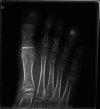
Figure 1
Figure 1
X-ray demonstrating the large nidus and the preservation of the proximal epiphysis. The important swelling of the fourth toe is well demonstrated.
Figure 2
Figure 2
T2-weighted image on MRI, showing the sclerotic nidus hyposignal and the surrounding inflammation of the soft tissues around (hypersignal).
Figure 3
Figure 3
Gadolinium-enhanced T1-weighted images on MRI, showing the relative avascular nidus surrounded by hypervascular inflamed tissues.
Figure 4
Figure 4
Histologic analysis demonstrating the characteristic pink-staining osteoid in a fibrovascular stroma (H&E staining, ×300).
Figure 5
Figure 5
(a) X-ray demonstrating the lyophilized bone graft three week after the intervention. (b) X-ray demonstrating the complete incorporation and remodeling of the graft one year after the intervention.

References

    1. Jaffe H. L. Osteoid-osteoma. Archives of Surgery. 1935;31(5):p. 709. doi: 10.1001/archsurg.1935.01180170034003. - DOI
    1. Hasegawa T., Hirose T., Sakamoto R., Seki K., Ikata T., Hizawa K. Mechanism of pain in osteoid osteomas: an immunohistochemical study. Histopathology. 1993;22(5):487–491. doi: 10.1111/j.1365-2559.1993.tb00163.x. - DOI - PubMed
    1. Ruggieri P., Angelini A., Montalti M., et al. Tumours and tumour-like lesions of the hip in the paediatric age: a review of the Rizzoli experience. Hip International. 2009;19(Supplement 6):S35–S45. doi: 10.1177/112070000901906s07. - DOI - PubMed
    1. Zampa V., Bargellini I., Ortori S., Faggioni L., Cioni R., Bartolozzi C. Osteoid osteoma in atypical locations: the added value of dynamic gadolinium-enhanced MR imaging. European Journal of Radiology. 2009;71(3):527–535. doi: 10.1016/j.ejrad.2008.05.010. - DOI - PubMed
    1. Mirra J. M., Kameda N., Rosen G., Eckardt J. Primary osteosarcoma of toe phalanx: first documented case. Review of osteosarcoma of short tubular bones. The American Journal of Surgical Pathology. 1988;12(4):300–307. doi: 10.1097/00000478-198804000-00007. - DOI - PubMed

Publication types

LinkOut - more resources