Novel Protocols for Scalable Production of High Quality Purified Small Extracellular Vesicles from Bovine Milk
- PMID: 34367882
- PMCID: PMC8342262
- DOI: 10.7150/ntno.62213
Novel Protocols for Scalable Production of High Quality Purified Small Extracellular Vesicles from Bovine Milk
Abstract
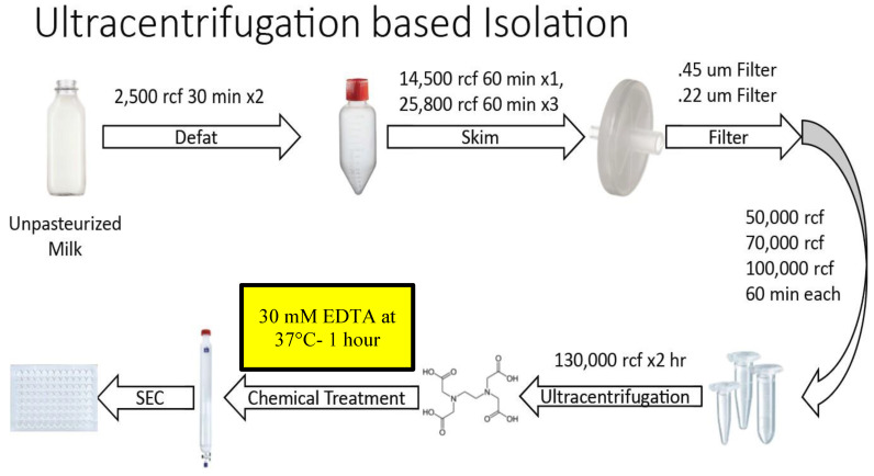
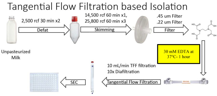
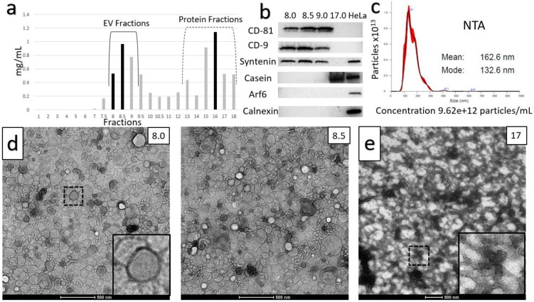
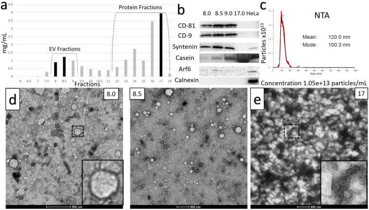
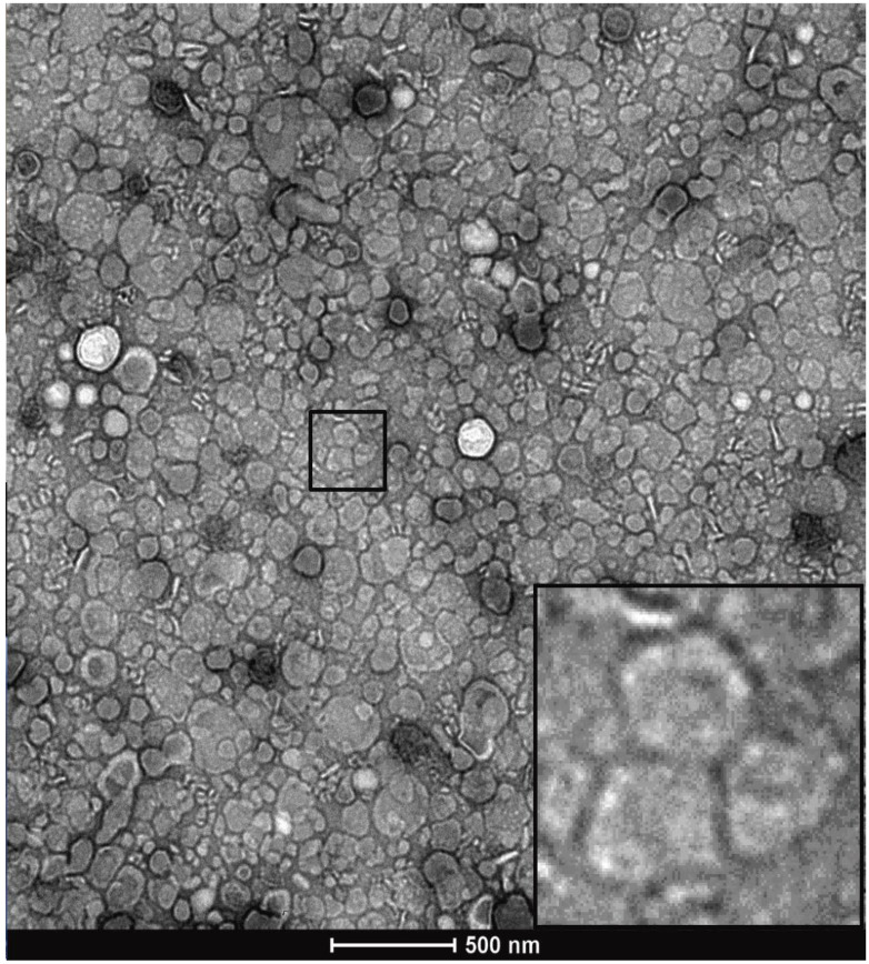
Extracellular Vesicles (EVs) are cell-secreted nanovesicles that have unique potential for encapsulating and targeting "difficult-to-drug" therapeutic cargos. Milk provides an enriched source of EVs, and of particular interest to the drug delivery field, small EVs. Small EVs are distinguished from large EVs by membrane components, biogenesis mechanism and downstream functionality - in particular, small EVs are primarily composed of exosomes, which show high stability in vivo and naturally function in the targeted delivery of biological materials to cells. Moreover, bovine milk is abundantly produced by the dairy industry, widely consumed, and generally well tolerated by humans. Importantly, there is evidence that milk exosomes and small EVs are efficiently taken up into the circulation from the gut, providing the opportunity for their use in administration of therapeutics such as microRNAs or peptides not typically available via an oral route. Unfortunately, present methods for isolation do not efficiently separate EVs from milk proteins, resulting in contamination that is not desirable in a clinical-grade therapeutic. Herein, we present novel EV purification methods focused on optimized timing and levels of temperature and divalent cation chelation. Incorporation of these solubilization steps into centrifugation- and tangential flow filtration-based methods provide large amounts of purified small EVs at ultra-dense concentrations, which are substantially free from contaminating milk proteins. Remarkably, these ultra-dense isolates equal 10 to 15% of the starting volume of milk indicating a prodigious rate of small EV production by mammary glands. Our approach enables gentle, scalable production of ultrastructurally and functionally intact small EVs from milk, providing a path to their industrial scale purification for oral delivery of therapeutic biologics and small drugs.
Keywords: Bovine Milk Extracellular Vesicles; Drug Delivery vehicle; Exosomes; Small Extracellular Vesicle Isolation; Small Extracellular Vesicles; Tangential Flow Filtration.
© The author(s).
Conflict of interest statement
Competing Interests: R.G.G. and S.R.M. are company officers at the Tiny Cargo Company Inc., which has licensed technology from Virginia Tech.
Figures
References
-
- Boulanger CM, Loyer X, Rautou PE, Amabile N. Extracellular vesicles in coronary artery disease. Nat Rev Cardiol. 2017;14:259–72. - PubMed
-
- Pegtel DM, Gould SJ. Exosomes. Annu Rev Biochem. 2019;88:487–514. - PubMed
-
- Thery C, Witwer KW, Aikawa E, Alcaraz MJ, Anderson JD, Andriantsitohaina R. et al. Minimal information for studies of extracellular vesicles 2018 (MISEV2018): a position statement of the International Society for Extracellular Vesicles and update of the MISEV2014 guidelines. J Extracell Vesicles. 2018;7:1535750. - PMC - PubMed
Publication types
MeSH terms
Substances
Grants and funding
LinkOut - more resources
Full Text Sources
Other Literature Sources