. 2021 Jul 22:11:705384.
doi: 10.3389/fonc.2021.705384. eCollection 2021.

Wnt/β-Catenin Inhibition Disrupts Carboplatin Resistance in Isogenic Models of Triple-Negative Breast Cancer

Affiliations

Wnt/β-Catenin Inhibition Disrupts Carboplatin Resistance in Isogenic Models of Triple-Negative Breast Cancer

Willy Antoni Abreu de Oliveira et al. Front Oncol. .

Abstract

Triple-Negative Breast Cancer (TNBC) is the most aggressive breast cancer subtype, characterized by limited treatment options and higher relapse rates than hormone-receptor-positive breast cancers. Chemotherapy remains the mainstay treatment for TNBC, and platinum salts have been explored as a therapeutic alternative in neo-adjuvant and metastatic settings. However, primary and acquired resistance to chemotherapy in general and platinum-based regimens specifically strongly hampers TNBC management. In this study, we used carboplatin-resistant in vivo patient-derived xenograft and isogenic TNBC cell-line models and detected enhanced Wnt/β-catenin activity correlating with an induced expression of stem cell markers in both resistant models. In accordance, the activation of canonical Wnt signaling in parental TNBC cell lines increases stem cell markers' expression, formation of tumorspheres and promotes carboplatin resistance. Finally, we prove that Wnt signaling inhibition resensitizes resistant models to carboplatin both in vitro and in vivo, suggesting the synergistic use of Wnt inhibitors and carboplatin as a therapeutic option in TNBC. Here we provide evidence for a prominent role of Wnt signaling in mediating resistance to carboplatin, and we establish that combinatorial targeting of Wnt signaling overcomes carboplatin resistance enhancing chemotherapeutic drug efficacy.

Keywords: WNT pathway; cancer stem cells; patient-derived xenograft models; platinum-resistance; triple negative breast cancer.


Conflict of interest statement

The authors declare that the research was conducted in the absence of any commercial or financial relationships that could be construed as a potential conflict of interest.

Figures
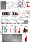
Figure 1
Figure 1
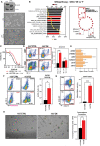
Carboplatin-resistant TNBC cells are characterized by enhanced WNT/β-catenin pathway activity, stem cell marker expression, and tumorsphere formation capacity. (A) Phase contrast microscope images of 468’P and 468’R cells. Scale bar: 100 μm. (B) Non-linear fit model of [CAR] μM vs normalized response for IC50 determination. (468’P: n=6, R2 = 0.92) (468’R: n=4, R2 = 0.95). (C) Growth curve and statistical analysis for 468’P and 468’R cells treated with VEH or CAR using two-way ANOVA. Statistical significance is reported for day 6 of treatment (n=4). (D) Representative flow cytometry scatterplots of annexin V staining of cells treated with 2 μM CAR for 72 hours (left) and respective statistical analysis (right) using multiple t-tests corrected for multiple comparisons with the Holms-Sidak method (n=3). (E) Enriched gene sets from Hallmarks and KEGG databases by one-tailed GSEA ranked by Normalized enrichment score (NES), illustrating pathways most significantly deregulated between 468’P and 468’R. (F) Enrichment map of one-tailed GSEA hits from Wikipathways database. Rectangles highlight clusters of gene sets with significant overlap and are labeled using AutoAnnotate on Cytoscape. (G) GSEA of hESCs (26) (left) and Cancer Progenitor (46) (right) gene sets in 468’R vs 468’P cells. (H) GSEA of NANOG, OCT4, and SOX2 target genes determined by ChIP-SEQ in hESC’s (26) in 468’R vs. 468’P cells. (I) Western blot of active non-phosphorylated β-catenin in 468’P and 468’R. β-actin was used as the loading control. (J) Representative scatterplots of flow cytometric analysis of aldehyde dehydrogenase activity (left). DEAB panels refer to internal controls in which ALDH activity is blocked with diethylaminobenzaldehyde to determine the background signal generated by unconverted ALDH substrate. TEST panels refer to the experimental samples where substrate for fluorimetric determination of ALDH activity is supplied. TEST samples are normalized to background fluorescence measured in DEAB internal controls and presented as the mean + standard error of the mean percentage of ALDH+ cells in 468’R (n=5) and 468’P (n=7) (right). Welch’s t-test. (K) Representative scatterplots of flow cytometric analysis of CD44-PE and CD24-APC immunolabeling (left) and corresponding statistical analysis of the mean percentage of CD44+/CD24- cells (right; n=3). Welch’s t-test. (L) qRT-PCR of Wnt target AXIN2 and stem cell markers in 468’R cells vs. 468’P (n=4). Multiple t-tests. (M) Representative brightfield images of tumorspheres generated from 468’P and 468’R cells (left, scale bar: 50 μm) and statistical analysis of mean tumorsphere forming units (number of spheres/number of seeded single cells) (right; n=3). Welch’s t-test. (Barplots represent mean + SEM. *p < 0.05; **p < 0.01; ****p < 0.0001; ns, non significant).
Figure 2
Figure 2
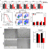
Pharmacological in vitro Wnt induction prevents carboplatin-induced apoptosis and upregulates stem cell marker expression. (A) Representative flow cytometry scatterplots (left) of Wnt-reporter MDA-MB-468 TOPGFP cells treated with vehicle (DMSO) or GSK3 inhibitor (CHIR, 4 μM) for 12 hours and statistical analysis of the mean frequency of GFP+ cells using Welch’s t-test (right, n=3). (B) Phase-contrast microscopy images of 468’P cells treated with or without carboplatin in the presence of CHIR or DMSO (left, scale bar: 100 μm) and statistical analysis of absolute cell numbers after 72 hours of each treatment using One-way ANOVA corrected for multiple comparisons using the Holm-Sidak method (n=4). (C) Representative flow cytometry scatterplots of annexin V staining of 468’P cells (left) treated with or without carboplatin in the presence of DMSO or CHIR (4 μM) for 72 hours and statistical analysis of the mean frequency of annexin V positive cells (right) using one-way ANOVA corrected for multiple comparisons using the Holm-Sidak method (n=3). (D) Relative mRNA expression of Wnt target and stem cell markers upon 72-hour treatment with DMSO or CHIR (4 μM) in 468’P cells (n=3). Multiple t-tests with Holm-Sidak correction for multiple comparisons. (Barplots represent mean + SEM. *p < 0.05; **p < 0.01; ***p < 0.001; ****p < 0.0001; ns, non significant).
Figure 3
Figure 3
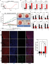
β-catenin overexpression in 468’P induces carboplatin-resistance, pluripotency-related gene expression, and cancer stem cell features. (A) Western blot (top) of total β-catenin in MDA-MB-468 cells transduced with an empty vector or truncated, constitutively active β-catenin isoform ΔN90 and phase-contrast microscopy (down). (B) Enriched gene sets from Wikipathways database by one-tailed GSEA of ranked DEGs between 468’OE and 468’P sorted by normalized enrichment score (left) and enrichment map illustrating pathways most significantly different between 468’OE and 468’P (right). (C) Non-linear fit model of [CAR] vs. normalized response for IC50 determination. (468’OE: n=6, R2 = 0.92; 468’CTRL: n=6, R2 = 0.95). (D) Representative flow cytometry scatterplots of annexin V staining (left) of 468’CTRL and 468’OE cells treated with carboplatin 2 μM for 72h and statistical analysis of the mean frequency of annexin V positive cells using one-way ANOVA corrected for multiple comparisons using the Holm-Sidak method (right, n=3). (E) mRNA level fold change (Log2) of CTNNB1 (β-catenin), Wnt target AXIN2, and stem cell markers in 468’OE cells vs. 468’CTRL (n=4). Multiple t-tests with Holms-Sidak correction for multiple comparisons. (F) Representative scatterplots of flow cytometric analysis of aldehyde dehydrogenase activity (left) and statistical analysis of the mean percentage of ALDH+ cells in 468’OE (n=5) and 468’CTRL (n=5) using Welch’s t-test (right). (G) Representative scatterplots of flow cytometric analysis of CD44-PE and CD24-APC immunolabeling (left) and corresponding statistical analysis of the mean percentage of CD44+/CD24- cells using Welch’s t-test (right; n=3). (H) Representative brightfield images of tumorspheres generated from 468’CTRL and 468’OE cells (left, scale bar: 50 μm) and statistical analysis of mean tumorsphere forming units (number of spheres/number of seeded single cells) using Welch’s t-test (right; n=3). (Barplots represent mean + SEM. *p < 0.05; **p < 0.01; ***p < 0.001; ****p < 0.0001; ns, non significant).
Figure 4
Figure 4
Wnt inhibitor LGK974 disrupts carboplatin resistance and pluripotency gene expression. (A) Absolute cell number of 468’R cells treated for 72 hours with increasing concentrations of LGK974 in the presence or absence of 2 μM carboplatin. Multiple t-tests (n=3). (B) Phase-contrast microscopy of 468’R cells treated for 72 hours with 200 nM LGK974 or DMSO in the presence or absence of 2 μM carboplatin (left). Mean frequency of annexin V positive cells in 468’P and 468’R cells treated with or without 200 nM LGK974 in presence or absence of 2 μM carboplatin showing the resensitization of 468’R cells to carboplatin when co-treated with Wnt inhibitor (right, n=3). One-way Anova with correction for multiple comparisons using the Holm-Sidak method. (C) Relative mRNA expression of Wnt target and stem cell markers upon 72-hour treatment with DMSO or 200 nM LGK974 with or without 2 μM carboplatin in 468’R cells (n=3). Multiple t-tests: Unt vs. Unt+LGK & CAR vs. CAR+LGK. (Barplots represent mean + SEM. *p < 0.05; **p < 0.01; ****p < 0.0001; ns, non significant).
Figure 5
Figure 5
Inducible β-catenin shRNA disrupts carboplatin-resistance and stem cell function in 468’R cells. (A) Relative mRNA expression level of CTNNB1 (β-catenin) and Wnt target and pluripotency markers in 468’R cells transduced with inducible CTNNB1-targeting or SCRMBL shRNAs, in the presence or absence of doxycycline (n=3). Welch’s t-test (Dox vs. no Dox). (B) Non-linear fit model of [CAR] vs normalized response for IC50 determination in iCTNNB1-KD cells in presence or absence of doxycyclin. (n=3, R2 + DOX: 0.93, R2 - DOX: 0.95). (C) Representative scatterplots of flow cytometric analysis of apoptosis by annexin V staining of 468’R iCTNNB1-KD and 468’R iSCRBML cells treated with or without 2 μM carboplatin for 72h in presence or absence of doxycycline (left) and corresponding statistical analysis of the mean frequency of annexin V positive cells (right, n=6). One-way Anova with correction for multiple comparisons using the Holm-Sidak method. (D) Representative brightfield images of tumorspheres generated from 468’R iSCRMBL and 468’R iCTNNB1-KD cells (left, scale bar: 50 μm) and statistical analysis of mean tumorsphere forming units (number of spheres/number of seeded single cells) (right; n=3). Welch’s t-test (Dox vs. no Dox). (Barplots represent mean + SEM. *p < 0.05; ***p < 0.001; ****p < 0.0001; ns, non significant).
Figure 6
Figure 6
WNT inhibition disrupts in vivo carboplatin-resistance in a carboplatin-resistant TNBC Patient-Derived Xenograft. (A) Tumor growth curves of carboplatin-sensitive BRC016 TNBC PDX model. Nine out of 10 mice show a complete response to treatment. One animal (red line) had a very delayed response and still had residual tumor mass after three weeks of treatment. The residual xenograft resumed growth after carboplatin treatment was stopped. This tumor was collected to establish a carboplatin-resistant model (C4O). (B) Comparative gene expression analysis by qRT-PCR of Wnt target AXIN2 and stem cell markers in BRC016 carboplatin-sensitive PDX and the C4O carboplatin-resistant isogenic PDX (n=4). Welch’s t-test. (C) Tumor growth curves of C4O carboplatin-resistant PDX treated with VEH, LGK974, CAR, or CAR+LGK showing reduced tumor growth in the combinatorial treatment arm (VEH, CAR, CAR+LGK n=6 and LGK n=5) (left). Two-way ANOVA with Tukey correction. The green-shadowed area under the curve represents highlights the time points in which the difference between CAR and CAR+LGK is statistically significant. Representative photographs of tumors in each treatment arm at day 21 of treatment (right). (D) mRNA level fold change (Log2) vs. VEH treatment (no carboplatin and no Wnt inhibitor) in tumors dissected at treatment endpoint (21 days) (n=3). Multiple t-tests (VEH vs. LGK and CAR vs. CAR+LGK). (E) Representative confocal microscopy images (left) of TUNEL staining in green and human pan-cytokeratin immunolabeling in red and respective quantification and statistical analysis of TUNEL positive cells (VEH n=4, LGK n=4, CAR & CAR+LGK n=6). One-way ANOVA with correction for multiple comparisons using the Holm-Sidak method. (Barplots represent mean + SEM. *p < 0.05; **p < 0.01; ****p < 0.0001; ns, non significant).
Figure 7
Figure 7
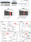
Wnt signaling is deregulated in patients with platinum-resistant TNBCs, high-grade serous ovarian cancer, and isogenic cisplatin-resistant ovarian cancer cell lines. (A) Summary of datasets and analysis methodology used. Functional enrichment and mapping as previously reported (40, 41, 60). (B) Enriched KEGG gene sets in patients with platinum-resistant TNBC (left) and HGSOC (right). (C) Enrichment maps for visualization of enriched KEGG gene sets in patients with platinum-resistant TNBC (left) and HGSOC (right). (D) Enrichment maps for visualization of gProfiler functional enrichment analysis of ranked, upregulated DEGs in patients with platinum-resistant TNBC (left) and HGSOC (right).

Similar articles

Cited by

References

    1. Carey L, Winer E, Viale G, Cameron D, Gianni L. Triple-Negative Breast Cancer: Disease Entity or Title of Convenience? Nat Rev Clin Oncol (2010) 7:683–92. 10.1038/nrclinonc.2010.154 - DOI - PubMed
    1. Marra A, Trapani D, Viale G, Criscitiello C, Curigliano G. Practical Classification of Triple-Negative Breast Cancer: Intratumoral Heterogeneity, Mechanisms of Drug Resistance, and Novel Therapies. NPJ Breast Cancer (2020) 6:1–16. 10.1038/s41523-020-00197-2 - DOI - PMC - PubMed
    1. Liedtke C, Mazouni C, Hess KR, André F, Tordai A, Mejia JA, et al. . Response to Neoadjuvant Therapy and Long-Term Survival in Patients With Triple-Negative Breast Cancer. J Clin Oncol (2008) 26:1275–81. 10.1200/JCO.2007.14.4147 - DOI - PubMed
    1. Rottenberg S, Disler C, Perego P. The Rediscovery of Platinum-Based Cancer Therapy. Nat Rev Cancer (2021) 21:37–50. 10.1038/s41568-020-00308-y - DOI - PubMed
    1. Turner NC, Reis-Filho JS, Russell AM, Springall RJ, Ryder K, Steele D, et al. . BRCA1 Dysfunction in Sporadic Basal-Like Breast Cancer. Oncogene (2007) 26:2126–32. 10.1038/sj.onc.1210014 - DOI - PubMed