Review
. 2021 Jul 23:9:707387.
doi: 10.3389/fchem.2021.707387. eCollection 2021.

Recent Advances in Mass Spectrometry-Based Glycomic and Glycoproteomic Studies of Pancreatic Diseases

Affiliations
Review

Recent Advances in Mass Spectrometry-Based Glycomic and Glycoproteomic Studies of Pancreatic Diseases

Dylan Nicholas Tabang et al. Front Chem. .

Abstract

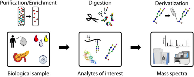
Modification of proteins by glycans plays a crucial role in mediating biological functions in both healthy and diseased states. Mass spectrometry (MS) has emerged as the most powerful tool for glycomic and glycoproteomic analyses advancing knowledge of many diseases. Such diseases include those of the pancreas which affect millions of people each year. In this review, recent advances in pancreatic disease research facilitated by MS-based glycomic and glycoproteomic studies will be examined with a focus on diabetes and pancreatic cancer. The last decade, and especially the last five years, has witnessed developments in both discovering new glycan or glycoprotein biomarkers and analyzing the links between glycans and disease pathology through MS-based studies. The strength of MS lies in the specificity and sensitivity of liquid chromatography-electrospray ionization MS for measuring a wide range of biomolecules from limited sample amounts from many sample types, greatly enhancing and accelerating the biomarker discovery process. Furthermore, imaging MS of glycans enabled by matrix-assisted laser desorption/ionization has proven useful in complementing histology and immunohistochemistry to monitor pancreatic disease progression. Advances in biological understanding and analytical techniques, as well as challenges and future directions for the field, will be discussed.

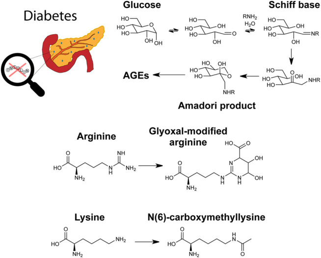
Keywords: diabetes; glycation; glycomics; glycoproteomics; glycosylation; mass spectrometry; pancreatic cancer; pancreatitis.


Conflict of interest statement

The authors declare that the research was conducted in the absence of any commercial or financial relationships that could be construed as a potential conflict of interest.

Figures

FIGURE 1
FIGURE 1
A representative workflow for mass spectrometry-based glycoproteomic analyses. Purification/enrichment, digestion, and derivatization of analytes may be performed at the glycoprotein, glycopeptide, and glycan levels.
FIGURE 2
FIGURE 2
Molecular modeling showing the docking of model glycopeptides (A, AcP(α-GalNAc)TLTH-NMe; (B), AcP(disialyl core 1)TLTH-NMe; (C), Ac-P(sialyl core 2)TLTH-NMe) with the mucin-selective protease StcE from E. coli. Reproduced from Malaker, S. A., Pedram, K., Ferracane, M. J., Bensing, B. A., Krishnan, V., Pett, C., et al. (2019). The mucin-selective protease StcE enables molecular and functional analysis of human cancer-associated mucins. Proc. Natl. Acad. Sci. U.S.A. 116, 7278–7287. doi: 10.1073/pnas.1813020116 under the PNAS license to publish.
FIGURE 3
FIGURE 3
MALDI-TOF-MS spectra of released and derivatized N-glycans from pancreatic duct (A) and cancer cells (B–E). Reproduced from Holst, S., Belo, A.I., Giovannetti, E., Van Die, I., and Wuhrer, M. (2017). Profiling of different pancreatic cancer cells used as models for metastatic behaviour shows large variation in their N-glycosylation. Sci. Rep. 7, 16623. doi: 10.1038/s41598-017-16811-6 under a Creative Commons Attribution 4.0 International License (http://creativecommons.org/licenses/by/4.0/).
FIGURE 4
FIGURE 4
Graphical depiction of diabetes, a glycation pathway relevant to hemoglobin, and the advanced glycation end products (AGEs) glyoxal-modified arginine and N(6)-carboxymethyllysine.
FIGURE 5
FIGURE 5
Chromatogram (A) and tandem mass spectra of glycopeptides (B), MS1 of glycoforms from glycosylation site I1; (C), MS2 of the peptide part; (D), MS2 of the glycan (N-acetylglucosamine)5(Hexose)6(Sialic acid)3) from human alpha-1-acid glycoprotein enriched from plasma. Reproduced from Keser, T., Tijardović, M., Gornik, I., Lukić, E., Lauc, G., Gornik, O., et al. (2021). High-throughput and site-specific N-glycosylation analysis of human alpha-1-acid glycoprotein offers a great potential for new biomarker discovery. Mol. Cell. Proteomics, 100044. doi: 10.1074/mcp.RA120.002433 under a Creative Commons CC-BY license (https://creativecommons.org/licenses/by/4.0/).
FIGURE 6
FIGURE 6
Graphical depiction of pancreatitis, pancreatic cancer, and several related O-glycans (with glycosidic linkages shown) investigated as biomarkers. R = peptide.
FIGURE 7
FIGURE 7
H&E stain (A) and MALDI-FT-ICR MS images of N-glycans (B), overlay of four glycans corresponding to necrotic tissue, adenocarcinoma, tumor margin, and adjacent non-tumor tissue; (C), glycans from necrotic tissue; (D), glycans from adenocarcinoma; (E), glycans from tumor margin; (F), glycans from adjacent non-tumor tissue) released from stage 3 pancreatic tumor tissue. Reproduced from McDowell, C. T., Klamer, Z., Hall, J., West, C. A., Wisniewski, L., Powers, T. W., et al. (2020). Imaging Mass Spectrometry and Lectin Analysis of N-Linked Glycans in Carbohydrate Antigen-Defined Pancreatic Cancer Tissues. Mol. Cell. Proteomics 20, 100012. doi: 10.1074/mcp.RA120.002256 under a Creative Commons CC-BY license (https://creativecommons.org/licenses/by/4.0/).
FIGURE 8
FIGURE 8
Top-down (A,C) and bottom-up tandem mass spectra (B,D) of an O-glycoform of an insulin chain from mouse pancreatic islets. Reproduced with permission from Yu, Q., Canales, A., Glover, M. S., Das, R., Shi, X., Liu, Y., et al. (2017). Targeted Mass Spectrometry Approach Enabled Discovery of O-Glycosylated Insulin and Related Signaling Peptides in Mouse and Human Pancreatic Islets. Anal. Chem. 89, 9184–9191. doi: 10.1021/acs.analchem.7b01926. Copyright © 2017 American Chemical Society.

Similar articles

Cited by

  • Characterization of Mesothelin Glycosylation in Pancreatic Cancer: Decreased Core Fucosylated Glycoforms in Pancreatic Cancer Patients' Sera.
    Duran A, Guerrero PE, Ortiz MR, Pérez Del Campo D, Castro E, Garcia-Velasco A, Fort E, de Llorens R, Saldova R, Llop E, Peracaula R. Duran A, et al. Biomedicines. 2022 Aug 10;10(8):1942. doi: 10.3390/biomedicines10081942. Biomedicines. 2022. PMID: 36009489 Free PMC article.
  • A Spin-Tip Enrichment Strategy for Simultaneous Analysis of N-Glycopeptides and Phosphopeptides from Human Pancreatic Tissues.
    Tabang DN, Wang D, Li L. Tabang DN, et al. J Vis Exp. 2022 May 4;(183):10.3791/63735. doi: 10.3791/63735. J Vis Exp. 2022. PMID: 35604151 Free PMC article.
  • Proteogenomic data and resources for pan-cancer analysis.
    Li Y, Dou Y, Da Veiga Leprevost F, Geffen Y, Calinawan AP, Aguet F, Akiyama Y, Anand S, Birger C, Cao S, Chaudhary R, Chilappagari P, Cieslik M, Colaprico A, Zhou DC, Day C, Domagalski MJ, Esai Selvan M, Fenyö D, Foltz SM, Francis A, Gonzalez-Robles T, Gümüş ZH, Heiman D, Holck M, Hong R, Hu Y, Jaehnig EJ, Ji J, Jiang W, Katsnelson L, Ketchum KA, Klein RJ, Lei JT, Liang WW, Liao Y, Lindgren CM, Ma W, Ma L, MacCoss MJ, Martins Rodrigues F, McKerrow W, Nguyen N, Oldroyd R, Pilozzi A, Pugliese P, Reva B, Rudnick P, Ruggles KV, Rykunov D, Savage SR, Schnaubelt M, Schraink T, Shi Z, Singhal D, Song X, Storrs E, Terekhanova NV, Thangudu RR, Thiagarajan M, Wang LB, Wang JM, Wang Y, Wen B, Wu Y, Wyczalkowski MA, Xin Y, Yao L, Yi X, Zhang H, Zhang Q, Zuhl M, Getz G, Ding L, Nesvizhskii AI, Wang P, Robles AI, Zhang B, Payne SH; Clinical Proteomic Tumor Analysis Consortium. Li Y, et al. Cancer Cell. 2023 Aug 14;41(8):1397-1406. doi: 10.1016/j.ccell.2023.06.009. Cancer Cell. 2023. PMID: 37582339 Free PMC article. Review.
  • Clinical glycoproteomics: methods and diseases.
    Wang Y, Lei K, Zhao L, Zhang Y. Wang Y, et al. MedComm (2020). 2024 Oct 4;5(10):e760. doi: 10.1002/mco2.760. eCollection 2024 Oct. MedComm (2020). 2024. PMID: 39372389 Free PMC article. Review.
  • Tetrazine bioorthogonal chemistry derived in vivo imaging.
    Zhao G, Li Z, Zhang R, Zhou L, Zhao H, Jiang H. Zhao G, et al. Front Mol Biosci. 2022 Nov 16;9:1055823. doi: 10.3389/fmolb.2022.1055823. eCollection 2022. Front Mol Biosci. 2022. PMID: 36465558 Free PMC article. Review.

References

    1. Abrahams J. L., Taherzadeh G., Jarvas G., Guttman A., Zhou Y., Campbell M. P. (2020). Recent Advances in Glycoinformatic Platforms for Glycomics and Glycoproteomics. Curr. Opin. Struct. Biol. 62, 56–69. 10.1016/j.sbi.2019.11.009 - DOI - PubMed
    1. Ahn J.-M., Kim B.-G., Yu M.-H., Lee I.-K., Cho J.-Y. (2010). Identification of Diabetic Nephropathy-Selective Proteins in Human Plasma by Multi-Lectin Affinity Chromatography and LC-MS/MS. Prot. Clin. Appl. 4, 644–653. 10.1002/prca.200900196 - DOI - PubMed
    1. Akella N. M., Ciraku L., Reginato M. J. (2019). Fueling the Fire: Emerging Role of the Hexosamine Biosynthetic Pathway in Cancer. BMC Biol. 17, 52. 10.1186/s12915-019-0671-3 - DOI - PMC - PubMed
    1. Akimoto Y., Nouso K., Kato H., Miyahara K., Dohi C., Morimoto Y., et al. (2015). Serum N-Glycan Profiles in Patients with Intraductal Papillary Mucinous Neoplasms of the Pancreas. Pancreatology 15, 432–438. 10.1016/j.pan.2015.05.470 - DOI - PubMed
    1. Almaraz R. T., Tian Y., Bhattarcharya R., Tan E., Chen S.-H., Dallas M. R., et al. (2012). Metabolic Flux Increases Glycoprotein Sialylation: Implications for Cell Adhesion and Cancer Metastasis. Mol. Cell Proteomics 11, 1–12. 10.1074/mcp.M112.017558 - DOI - PMC - PubMed

LinkOut - more resources