Review
. 2021 Jul 22:9:681123.
doi: 10.3389/fcell.2021.681123. eCollection 2021.

Meiotic Crossover Patterning

Affiliations
Review

Meiotic Crossover Patterning

Nila M Pazhayam et al. Front Cell Dev Biol. .

Abstract

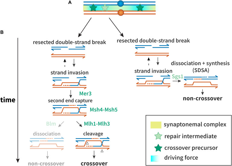
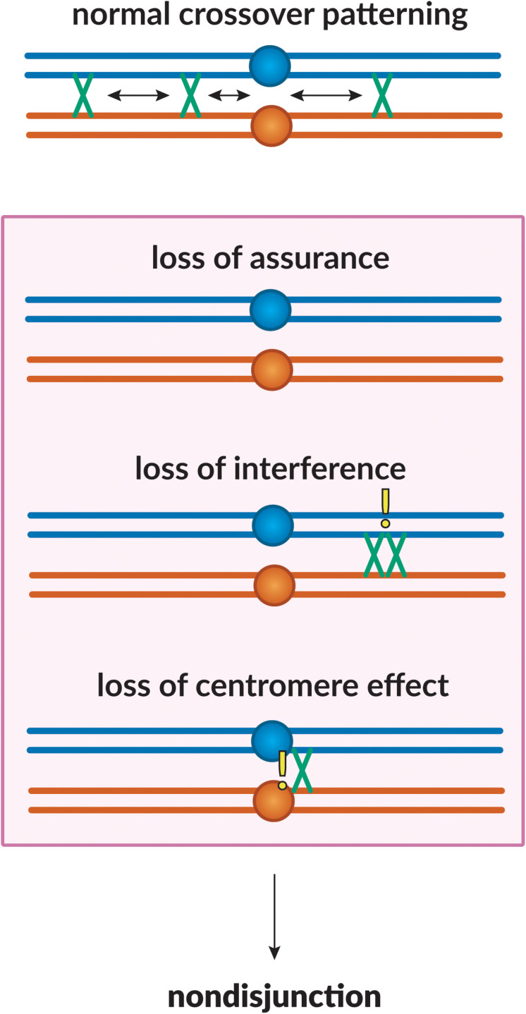
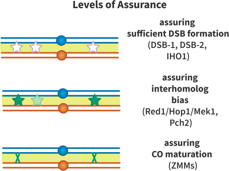
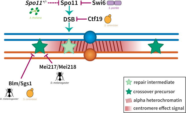
Proper number and placement of meiotic crossovers is vital to chromosome segregation, with failures in normal crossover distribution often resulting in aneuploidy and infertility. Meiotic crossovers are formed via homologous repair of programmed double-strand breaks (DSBs). Although DSBs occur throughout the genome, crossover placement is intricately patterned, as observed first in early genetic studies by Muller and Sturtevant. Three types of patterning events have been identified. Interference, first described by Sturtevant in 1915, is a phenomenon in which crossovers on the same chromosome do not occur near one another. Assurance, initially identified by Owen in 1949, describes the phenomenon in which a minimum of one crossover is formed per chromosome pair. Suppression, first observed by Beadle in 1932, dictates that crossovers do not occur in regions surrounding the centromere and telomeres. The mechanisms behind crossover patterning remain largely unknown, and key players appear to act at all scales, from the DNA level to inter-chromosome interactions. There is also considerable overlap between the known players that drive each patterning phenomenon. In this review we discuss the history of studies of crossover patterning, developments in methods used in the field, and our current understanding of the interplay between patterning phenomena.

Keywords: centromere; crossover assurance; interference; meiosis; recombination.


Conflict of interest statement

The authors declare that the research was conducted in the absence of any commercial or financial relationships that could be construed as a potential conflict of interest.

Figures

FIGURE 1
FIGURE 1
Proteins and mechanisms of crossover interference. (A) A driving force (teal), potentially protein aggregation or mechanical stress, results in designation of crossover precursors (stars, dark green) at spaced intervals. Repair intermediates that do not experience sufficient driving force (stars, light green) are not so designated, and become non-crossovers. (B) Interference at the DNA level. Crossover precursors are processed through the class I crossover pathway, involving the ZMM proteins. Mer3 promotes second end capture, and Msh4–Msh5 clamp to and stabilize branched molecules to prevent their dissociation and to recruit Msh1–3. Intermediates are resolved by Mlh1–Mlh3 to generate crossovers. Non-crossovers in yeast are thought to be exclusively processed through the synthesis-dependent strand annealing (SDSA) pathway, promoted by Sgs1. In other organisms, the Sgs1 ortholog Blm may also promote formation of non-crossovers after second end capture.
FIGURE 2
FIGURE 2
Crossover patterning phenomena. The proper placement of crossovers along the chromosome is governed by three patterning phenomena. Homologous chromosomes are shown in blue and orange, with crossovers between them shown in green. Loss of assurance results in a lack of crossing-over between a pair of homologs; loss of interference results in two crossovers being placed in close proximity to one another; loss of the centromere effect results in centromere-proximal crossovers. These phenomena can lead to a failure in proper chromosome segregation, leading to non-disjunction.
FIGURE 3
FIGURE 3
Chromatid interference. Each panel illustrates two crossovers on one bivalent, with each line representing one double-stranded DNA chromatid. The first (leftmost) crossover is the same in all cases, occurring between the two inner chromatids. (A) A 2-chromatid DCO. (B) Two possible 3-chromatid DCO configurations. (C) A 4-chromatid DCO. If there is no chromatid interference, the ratio of 2:3:4 should be 1:2:1.
FIGURE 4
FIGURE 4
Levels of crossover assurance. Assurance is enforced at three levels. DSB-1 and DSB-2 form a checkpoint in C. elegans that ensures that sufficient double-strand breaks (DSBs, white stars) are formed to generate an obligate crossover. In mouse, IHO1 plays a similar role. From this pool of DSBs, inter-homolog recombination is promoted by Red1, Hop1, Mek1, and Pch2 to ensure that recombination intermediates (light green stars) engage with the homologous chromosome to form crossover precursors (dark green stars). The driving force to generate crossover precursors may be aggregation of ZMM proteins at recombination nodules or mechanical stress that must be relieved by crossover formation. Efficient crossover maturation (dark green X) is ensured by the ZMM proteins that define the class I crossover pathway.
FIGURE 5
FIGURE 5
Suppression of crossovers near the centromere. The exclusion of crossovers near the centromere seems to be enforced at two levels. Crossover suppression in the highly repetitive alpha heterochromatin has been shown to be a result of the prevention of DSBs either through local chromatin organization (in S. cerevisiae) or the inactivation of Spo11 (in S. pombe and A. thaliana). Crossover suppression in the less repetitive beta heterochromatin and the proximal euchromatin has been shown to be regulated through Bloom syndrome helicase/Sgs1 (in D. melanogaster and S. cerevisiae) and dependent on distance from the centromere (represented by a centromere effect signal shown in red). Homologous chromosomes are shown in blue and orange with dark red lines in the pericentromeric region representing alpha heterochromatin. Light and dark green stars represent repair intermediates and crossover precursors, respectively.

References

    1. Agostinho A., Meier B., Sonneville R., Jagut M., Woglar A., Blow J., et al. (2013). Combinatorial regulation of meiotic Holliday junction resolution in C. elegans by HIM-6 (BLM) helicase, SLX-4, and the SLX-1, MUS-81 and XPF-1 nucleases. PLoS Genet. 9:e1003591. 10.1371/journal.pgen.1003591 - DOI - PMC - PubMed
    1. Ahuja J. S., Sandhu R., Mainpal R., Lawson C., Henley H., Hunt P. A., et al. (2017). Control of meiotic pairing and recombination by chromosomally tethered 26S proteasome. Science 355 408–411. 10.1126/science.aaf4778 - DOI - PMC - PubMed
    1. Albertson D. G., Thomson J. N. (1993). Segregation of holocentric chromosomes at meiosis in the nematode, Caenorhabditis elegans. Chromosome Res. 1 15–26. 10.1007/bf00710603 - DOI - PubMed
    1. Allers T., Lichten M. (2001). Differential timing and control of noncrossover and crossover recombination during meiosis. Cell 106 47–57. 10.1016/s0092-8674(01)00416-0 - DOI - PubMed
    1. Andersen S. L., Sekelsky J. (2010). Meiotic versus mitotic recombination: two different routes for double-strand break repair: the different functions of meiotic versus mitotic DSB repair are reflected in different pathway usage and different outcomes. Bioessays 32 1058–1066. 10.1002/bies.201000087 - DOI - PMC - PubMed

LinkOut - more resources