

The phenomenon of clasmatodendrosis
- PMID: 34368479
- PMCID: PMC8326353
- DOI: 10.1016/j.heliyon.2021.e07605
The phenomenon of clasmatodendrosis
Abstract
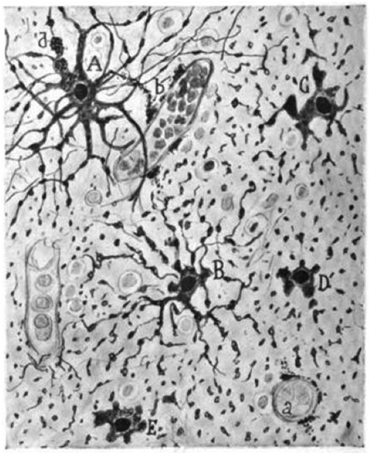
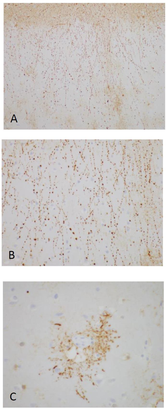
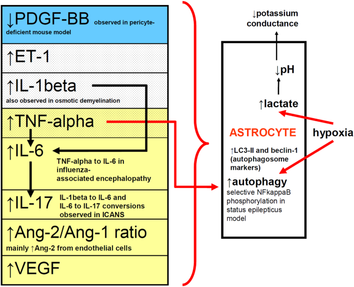
Clasmatodendrosis derives from the Greek for fragment (klasma), tree (dendron), and condition (- osis). Cajal first used the term in 1913: he observed disintegration of the distal cell processes of astrocytes, along with a fragmentation or beading of proximal processes closer to the astrocyte cell body. In contemporary clinical and experimental reports, clasmatodendrosis has been observed in models of cerebral ischemia and seizures (including status epilepticus), in elderly brains, in white matter disease, in hippocampal models and cell cultures associated with amyloid plaques, in head trauma, toxic exposures, demyelinating diseases, encephalitides and infection-associated encephalopathies, and in the treatment of cancer using immune effector cells. We examine evidence to support a claim that clasmatodendrotic astrocyte cell processes overtly bead (truncate) as a morphological sign of ongoing damage premortem. In grey and white matter and often in relationship to vascular lumina, beading becomes apparent with immunohistochemical staining of glial fibrillary acidic protein when specimens are examined at reasonably high magnification, but demonstration of distal astrocytic loss of processes may require additional marker study and imaging. Proposed mechanisms for clasmatodendrotic change have examined hypoxic-ischemic, osmotic-demyelinating, and autophagic models. In these models as well as in neuropathological reports, parenchymal swelling, vessel-wall leakage, or disturbed clearance of toxins can occur in association with clasmatodendrosis. Clasmatodendrotic features may serve as a marker for gliovascular dysregulation either acutely or chronically. We review correlative evidence for blood-brain barrier (BBB) dysfunction associated with astrocytic structural change, with attention to interactions between endothelial cells, pericytes, and astrocytic endfeet.
Keywords: Astrocyte; Blood-brain barrier; Clasmatodendrosis; Endfoot; Endothelial cell; Pericyte.
© 2021 The Authors.
Conflict of interest statement
The authors declare the following conflict of interests: Dr. Shamik Bhattacharyya receives personal fees from Alexion Pharmaceuticals and honoraria from UpToDate and Springer. Dr. Matthew Torre is supported by the 10.13039/100000054National Cancer Institute of the 10.13039/100000002National Institutes of Health under award number F32CA257210. The content is solely the responsibility of the authors and does not necessarily represent the official views of the National Institutes of Health.
Figures
References
-
- Ramón y., Cajal S. Contribución al conocimiento de la neuroglía del cerebro humano. Trabajos del Laboratorio de Investigaciones Biológicas de la Universidad de Madrid. 1913;11:255–315.
-
- Alzheimer A. Beiträge zur kenntnis der pathologischen neuroglia und ihrer beziehungen zu den abbauvorgangen im nervengewebe. In: Nissl F., Fischer G., editors. Vol. 3. 1910. pp. 410–562. (Histologische und histopathologische Arbeiten über die Grosshirnrinde mit besonderer Berücksichtung der pathologischen Anatomie der Geisteskrankheiten).
-
- Rosental V.S. Experimentelle studien über amöboide umwandlung der neuroglia. In: Nissl F., Alzheimer A., editors. Vol. 6. 1913. pp. 89–160. (Histologische und histopathologische Arbeiten über die Grosshirnrinde mit besonderer Berücksichtung der pathologischen Anatomie der Geisteskrankheiten).
-
- Del Río-Hortega P. Noticia de un nuevo y fácil método para la coloración de la neuroglía y del tejido conjuntivo. Trabajos del Laboratorio de Investigaciones Biológicas de la Universidad de Madrid. 1917;15:367–378.
Publication types
Grants and funding
LinkOut - more resources
Full Text Sources
