. 2021 Jun:22:e00132.
doi: 10.1016/j.bprint.2021.e00132.

3D Bioprinting for In Vitro Models of Oral Cancer: Toward Development and Validation

Affiliations

3D Bioprinting for In Vitro Models of Oral Cancer: Toward Development and Validation

Thafar Almela et al. Bioprinting. 2021 Jun.

Abstract

The tumor microenvironment (TME) of oral carcinomas has highly complex contents and a dynamic nature which is difficult to study using oversimplified two-dimensional (2D) cell culture systems. By contrast, three dimensional (3D) in vitro models such as spheroids, organoids, and scaffold-based constructs have been able to replicate tumors three-dimensionality and have allowed a better understanding of the role of various microenvironmental cues in the initiation and progression of cancer. However, the heterogeneity of TME cannot be fully reproduced by these traditional tissue engineering strategies since they are unable to control the organization of multiple cell types in a complex architecture. 3D bioprinting is an emerging field that can be leveraged to produce biomimetic and complex tissue structures. Bioprinting allows for controllable and precise placement of multicomponent bioinks composed of multiple biomaterials, different types of cells, and soluble factors according to the natural compartments of the target tissue, aiming to reproduce the equivalent of the complex tissue. As such, 3D bioprinting provides a unique opportunity to fabricate in vitro tumor models with a complexity similar to that of the in vivo oral carcinoma. This will facilitate a thorough investigation of cellular physiology, cancer progression, and anti-cancer drug screening with unprecedented control and reproducibility. In this review, we discuss the role of 3D bioprinting in reconstituting oral cancer, the prospects of application to fill the literature gap, and the challenges that need to be addressed in order to exploit this emerging technology for future work in oral cancer research.

Keywords: Oral cancer; Oral squamous cell carcinoma; Three-dimensional bioprinting; Three-dimensional tumor models; Tissue engineering.


Conflict of interest statement

Competing interests The authors declare no competing interests.

Figures

Figure 1.
Figure 1.. The last release of the GLOBOCAN 2020 data produced by the International Agency for Research on Cancer (IARC) regarding oral cavity and oropharyngeal cancers.
(A) estimated number of new cases of lip and oral cavity cancer; (B) estimated number of deaths in lip and oral cavity cancer; (C) estimated number of new cases of oropharynx cancer; (D) estimated number of deaths in oropharynx cancer. The data represents estimates of the incidence and mortality of oral cancer and Oropharynx for both sexes and all ages in 185 countries of the world. The incidence and mortality for each sex were estimated with an age-standardized rate (ASR) per 100,000. Data source: (GLOBOCAN) 2020, (https://gco.iarc.fr) [7].
Figure 2.
Figure 2.. Schematic diagrams of 3D bioprinting technologies.
Laser-induced forward tranfer bioprinting use lasers focused on a laser energy absorbing layer to generate a high gas pressure propelling the bioink onto a collector substrate. Thermal inkjet printers electrically heat the printhead to produce air-pressure pulses that expels droplets from the nozzle, whereas in piezoelectric inkjet printing no heating is used, but a direct mechanical pulse is applied to the fluid by a piezoelectric actuator that forces the bioink through the nozzle. Robotic dispensing or extrusion bioprinting use either pneumatic, piston- or screw-driven dispensing systems to extrude continuous beads of bioink on a building platform. Reproduced with permission from ref. [54].
Figure 3.
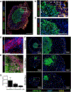
Figure 3.. Three-Dimensional Bioprinted Model of Pancreatic Cancer.
(A) Bioprinted tissue containing a patient-derived xenograft (PDX)-derived cell line from a human pancreatic cancer cell line surrounded by normal human primary pancreatic stellate cells (PSCs) and HUVECs after 7 days in culture. IF for epithelial cancer cells KRT8/18 (green), stromal fibroblasts VIM (red), and endothelial cells CD31 (yellow) with DAPI (blue) nuclear counterstain (Scale bar 500 μm). (B) The tumor-stromal border with VIM-positive cells within the tumor core, and KRT8/18 cells in the stroma (Scale bar 200 μm). (C) IF for KRT8/18 (green) and Ki67 (red) with DAPI (blue) (Scale bar 200 μm). (D) IF for α-SMA in PSCs after 7 days in culture (Scale bar 50 μm). (E) Digital image on bioprinted pancreatic tumor tissue printed with PSCs and HUVECs in the stromal compartment and human pancreatic cancer cell line (HPAFIIs) and HUVECs in the cancer compartment after 10 days of culture (Scale bar 100 μm). (F) Pancreatic tumor tissues printed as in (E) were treated for 6 days with either 10 or 50 μM gemcitabine. IF performed for KRT8/18 (green) and CD31 (yellow) with DAPI (blue). (G) Day 10 quantitation of total flux for bioprinted tissues grown as in (E). (IF: immunofluorescence; KRT8/18: Cytokeratin 8/18; VIM: vimentin; α-SMA: Alpha-Smooth muscle actin). Reproduced with permission from ref.[60].
Figure 4.
Figure 4.. Control of biochemical factors distribution in a bioprinted model.
Structural designs of 3D printed scaffold laden with DPSCs and spiked with BMP-2 and VEGF. Group 1: DPSCs printed structure using 2% collagen. Group 2: DPSC/BMP-2 printed structure using 2% collagen. Group 3: DPSC/dual growth factors of BMP-2 and VEGF using 2% collagen and 10% alginate/10% gelatin blend (DPSCs: Mesenchymal dental pulp derived stem cells; BMP-2: bone morphogenetic protein 2; VEGF: vascular endothelial growth factor). Reproduced with permission from ref.[63].
Figure 5.
Figure 5.. Bioprinting of vascular constructs.
(A1) Representative images of bioprinted fibroblast tubes made of 2% alginate loaded with 5×10⁶ cells/ml; (A2) left inset: different views of a printed Y-shaped cellular tube bioprinted using 2% sodium alginate solution, and right inset: dyed cells in blue and dyed living cells in green (reproduced with permission from Ref.[78]); (B) Direct cell patterning in collagen hydrogels using a near-infrared femtosecond laser. The confocal microscopy images show tube formation after 14 days in the YZ plane and aligned endothelial cells in the XY plane (reproduced with permission from Ref.[79]). (C) Scanning electron microscopy images showing a complex vascular network in poly(ethylene glycol) diacrylate hydrogel (Reproduced with permission from ref.[80]).
Figure 6.
Figure 6.. 3D bioprinting of the prevasularized tissue constructs.
(1) (A) Schematic of the bioprinting platform. (B) Bioprinted acellular construct featuring intended channels with gradient widths. (C) Bioprinted cellular construct with HUVECs encapsulated in the channels. (D–F) Fluorescent images demonstrating the bioprinting of heterogeneous cell-laden tissue constructs with uniform channel width. HUVECs (red) are encapsulated in the intended channels and HepG2 (green) are encapsulated in the surrounding area. (G–I) Fluorescent images demonstrating the bioprinting of heterogeneous cell-laden tissue constructs with gradient channel widths. Scale bars 250 μm. (2) Endothelial network formation after 1-week culture of the prevascularized tissue construct in vitro, (A–C) Confocal microscopy images show HUVECs (Green, CD31-positive) and supportive mesenchymal cells (10T1/2, Purple, alpha-smooth muscle actin (α-SMA)-positive) aligned within the patterned channel regions with different vessel sizes. (D) Cross-section view shows the endothelial cells (CD31-positive) form lumen-like structures along the bioprinted channels. (E) 3D view of the endothelial cells lining along the printed microchannel walls by confocal microscopy. Endothelial cells were labeled by fluorescent cell tracker (red) and stained by CD31 (green). Reproduced with permission from ref. [81].

References

    1. Warnakulasuriya S, Greenspan JS., Textbook of oral cancer: Prevention, diagnosis and management. Cham: Springer International Publishing, 2020. ISBN-10: 3030323153.
    1. Bray F, Ferlay J, Soerjomataram I, Siegel RL, Torre LA, Jemal A, Global cancer statistics 2018: Globocan estimates of incidence and mortality worldwide for 36 cancers in 185 countries, CA Cancer J Clin. 68 (2018) 394–424. 10.3322/caac.21492. - DOI - PubMed
    1. Siegel RL, Miller KD, Jemal A, Cancer statistics, 2020. CA Cancer J Clin. 70 (2020) 7–30. 10.3322/caac.21590. - DOI - PubMed
    1. Shield KD, Ferlay J, Jemal A, Sankaranarayanan R, Chaturvedi AK, Bray F, Soerjomataram I, The global incidence of lip, oral cavity, and pharyngeal cancers by subsite in 2012. CA Cancer J Clin. 67(2017) 51–64. 10.3322/caac.21384. - DOI - PubMed
    1. Yang Y-H, Warnakulasuriya S, Effect of comorbidities on the management and prognosis in patients with oral cancer. Translational Research in Oral Oncology. 1(2016) 1–8. 10.1177/2057178X16669961. - DOI