. 2021 Jul 26;6(7):567-580.
doi: 10.1016/j.jacbts.2021.05.004. eCollection 2021 Jul.

Implications of Iron Deficiency in STEMI Patients and in a Murine Model of Myocardial Infarction

Affiliations

Implications of Iron Deficiency in STEMI Patients and in a Murine Model of Myocardial Infarction

Javier Inserte et al. JACC Basic Transl Sci. .

Abstract

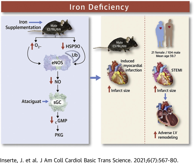
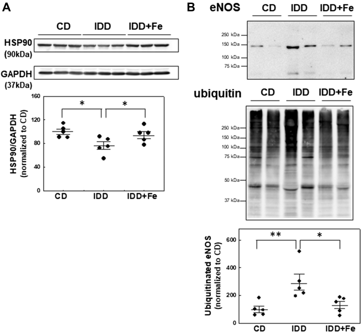
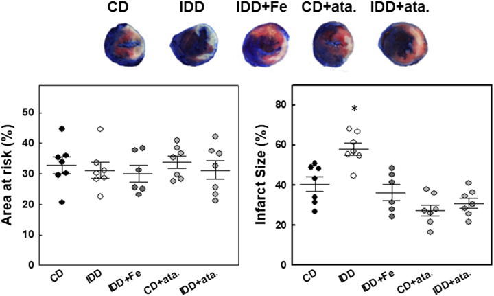
In patients with a first anterior ST-segment elevation myocardial infarction treated with primary percutaneous coronary intervention, iron deficiency (ID) was associated with larger infarcts, more extensive microvascular obstruction, and higher frequency of adverse left ventricular remodeling as assessed by cardiac magnetic resonance imaging. In mice, an ID diet reduced the activity of the endothelial nitric oxide synthase/soluble guanylate cyclase/protein kinase G pathway in association with oxidative/nitrosative stress and increased infarct size after transient coronary occlusion. Iron supplementation or administration of an sGC activator before ischemia prevented the effects of the ID diet in mice. Not only iron excess, but also ID, may have deleterious effects in the setting of ischemia and reperfusion.

Keywords: CK-MB, creatine kinase-myocardial band; CMR, cardiac magnetic resonance; HSP90, heat-shock protein 90; ID, iron deficiency; LV, left ventricular; MVO, microvascular obstruction; PKG, protein kinase G; STEMI, ST-segment elevation acute myocardial infarction; STIR, short tau inversion recovery; VASP, vasodilator-stimulated phosphoprotein; acute myocardial infarction; eNOS, endothelial nitric oxide synthase; endothelial nitric oxide synthase; iNOS, inducible nitric oxide synthase; iron deficiency; myocardial reperfusion; sGC, soluble guanylyl cyclase; soluble guanylate cyclase.


Conflict of interest statement

This study was funded by Instituto de Salud Carlos III, Spain, through the project PI16/00232 and the research network CIBERCV (CB16/11/00479), both cofunded by European Regional Development Fund, and by the Sociedad Española de Cardiología (Proyecto de Investigación Traslacional en Cardiología 2016). The authors have reported that they have no relationships relevant to the contents of this paper to disclose.

Figures

None
Graphical abstract
Figure 1
Figure 1
Patient Flowchart CMR = cardiac magnetic resonance.
Figure 2
Figure 2
ID Inhibits the eNOS/cGMP/PKG Pathway Representative Western blot and quantification of (A) total eNOS and its phosphorylated form and (B) the sodium dodecyl sulfate–resistant eNOS dimer in cardiac lysates obtained from the different experimental groups, n = 5 per group. (C) Representative Western blot and quantification of total eNOS and its dimeric form in isolated cardiomyocytes treated with deferasirox (DFX), n = 4 per group. (D) Quantification of myocardial content of nitrate/nitrite (NOx) and cGMP, n = 5 per group. (E) protein kinase G (PKG) activity evaluated by Western blot as PKG-dependent phosphorylation of its substrate vasodilator-stimulated phosphoprotein (VASP), n = 5 per group. (F) Representative Western blot of inducible nitric oxide synthase (iNOS). LPS corresponds to a mouse spleen sample obtained 6 h after lipopolysaccharide injection (10 mg/kg intraperitoneally). ∗P < 0.05, ∗∗P < 0.01 vs IDD group. CD = control diet; eNOS = endothelial nitric oxide synthase; IDD = iron-deficient diet; IDD+Fe = IDD plus iron supplementation. Data are mean ± SEM.
Figure 3
Figure 3
ID Increases Oxidative/Nitrosative Stress by Reducing Myocardial Antioxidant Capacity (A) Quantification of markers of oxidative (8-isoprostane and protein carbonyl) and nitrosative (nitrotyrosine) stress, n = 5 per group. (B) Representative ventricular cross-sections incubated with dihydroethidium (DHE). DHE fluorescence is expressed as arbitrary units, n = 4 per group. (C) Quantification of total antioxidant capacity and superoxide dismutase-1 (SOD1) activity, n =5 per group. (D) Time course of H2O2 production from isolated mitochondria after the addition of malate plus glutamate or succinate. F/F0 corresponds to arbitrary fluorescence values normalized with respect to baseline (excitation at 320 nm, emission at 440 nm), n = 3 per group. Data are mean ± SEM. ∗P < 0.05, ∗∗P < 0.01 vs IDD groups. Abbreviations as in Figure 2.
Figure 4
Figure 4
ID Reduces HSP90 Levels and Increases the Extent of eNOS Ubiquitination (A) Representative Western blot and quantification of HSP90. (B) Polyubiquitination levels of eNOS in cardiac lysates analyzed by using agarose-tandem ubiquitin-binding entities pull-downs. eNOS bands were normalized to their corresponding ubiquitin smears and ratios were normalized to ubiquitinated eNOS level in control samples. Representative Western blots are shown in all panels. Data are mean ± SEM. ∗P < 0.05, ∗∗P < 0.01 vs IDD group. Abbreviations as in Figure 2.
Figure 5
Figure 5
ID Increases Myocardial Susceptibility to Ischemia/Reperfusion Injury Area at risk, expressed as a percentage of left ventricular mass, and infarct size, measured by triphenyltetrazolium chloride staining and expressed as a percentage of the area at risk, in mouse hearts after 24-h of reperfusion in the different experimental groups. Representative examples are shown. Data are mean ± SEM. n = 6-7 per group. ∗P < 0.05 vs the CD group. Ata = ataciguat; other abbreviations as in Figure 2.

References

    1. Anker S.D., Comin Colet J., Filippatos G. Ferric carboxymaltose in patients with heart failure and iron deficiency. N Engl J Med. 2009;361:2436–2438. - PubMed
    1. Rocha B.M.L., Cunha G.J.L., Menezes Falcão L.F. The burden of iron deficiency in heart failure: therapeutic approach. J Am Coll Cardiol. 2018;71:782–793. - PubMed
    1. Meroño O., Cladellas M., Ribas-Barquet N. Iron deficiency is a determinant of functional capacity and health-related quality of life 30 days after an acute coronary syndrome. Rev Esp Cardiol. 2017;70:363–370. - PubMed
    1. Zeller T., Waldeyer C., Ojeda F. Adverse outcome prediction of iron deficiency in patients with acute coronary syndrome. Biomolecules. 2018;8:60. - PMC - PubMed
    1. González-D'Gregorio J., Miñana G., Núñez J. Iron deficiency and long-term mortality in elderly patients with acute coronary syndrome. Biomark Med. 2018;12:987–999. - PubMed