. 2021 Jul 10;24(8):102837.
doi: 10.1016/j.isci.2021.102837. eCollection 2021 Aug 20.

Newcastle-disease-virus-induced ferroptosis through nutrient deprivation and ferritinophagy in tumor cells

Affiliations

Newcastle-disease-virus-induced ferroptosis through nutrient deprivation and ferritinophagy in tumor cells

Xianjin Kan et al. iScience. .

Abstract

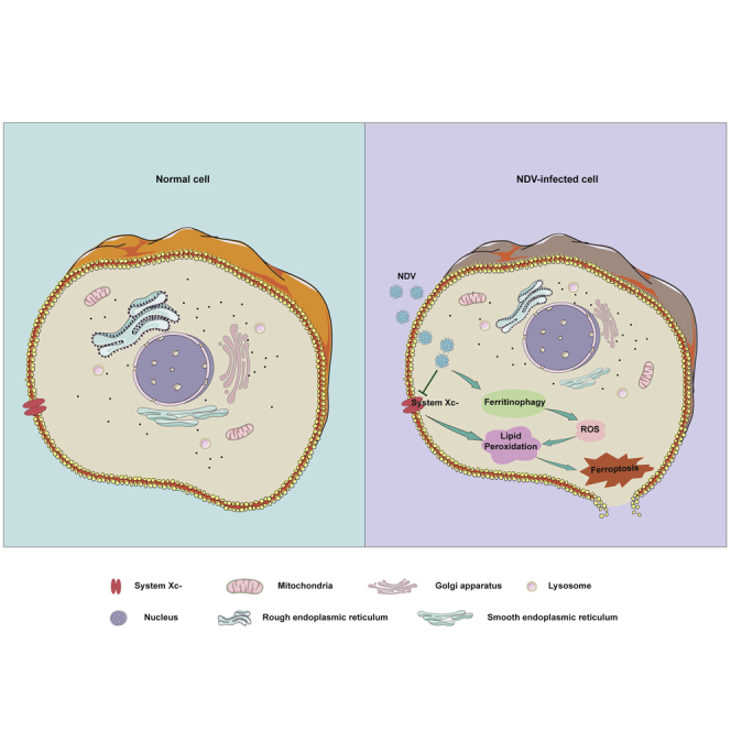
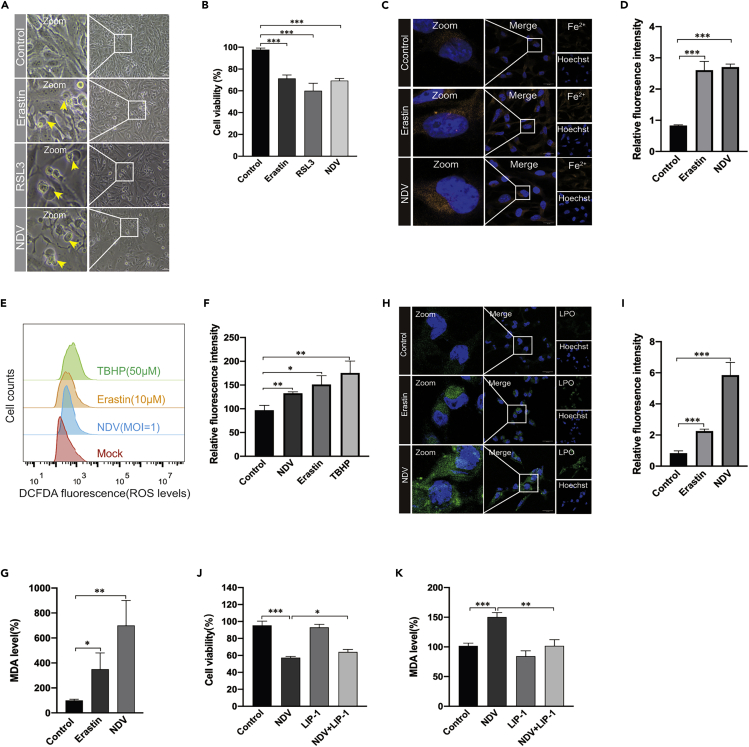
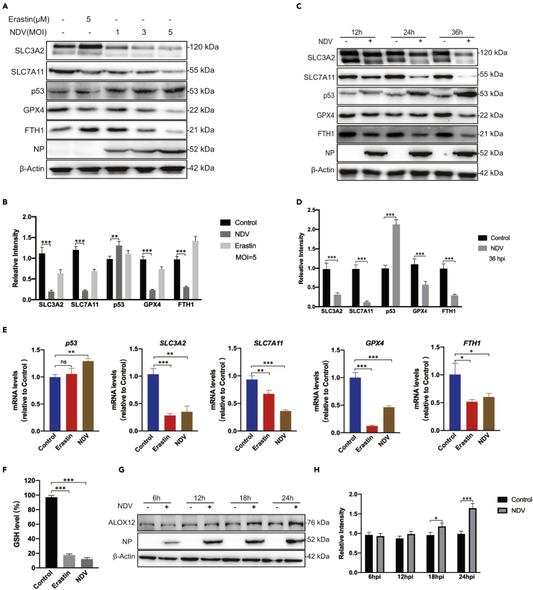
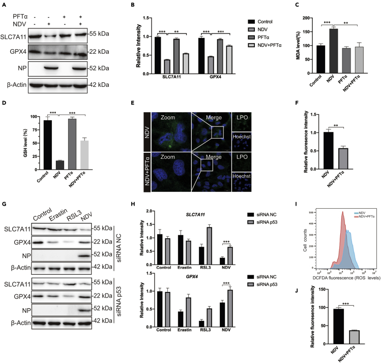
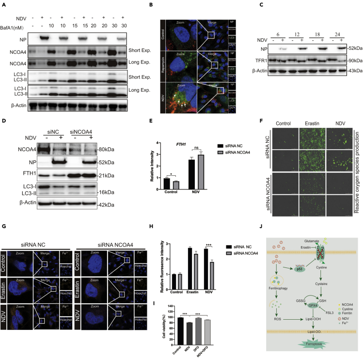
A number of new cell death processes have been discovered in recent years, including ferroptosis, which is characterized by the accumulation of lipid peroxidation products derived from iron metabolism. The evidence suggests that ferroptosis has a tumor-suppressor function. However, the mechanism by which ferroptosis mediates the response of tumor cells to oncolytic viruses remains poorly understood. The Newcastle disease virus (NDV) can selectively replicate in tumor cells. We show that NDV-induced ferroptosis acts through p53-SLC7A11-GPX4 pathway. Meanwhile, the levels of intracellular reactive oxygen species and lipid peroxides increased in tumor cells. Ferritinophagy was induced by NDV promotion of ferroptosis through the release of ferrous iron and an enhanced Fenton reaction. Collectively, these observations demonstrated that the NDV can kill tumor cells through ferroptosis. Our study provides novel insights into the mechanisms of NDV-induced ferroptosis and highlights the critical role of viruses in treating therapy-resistant cancers.

Keywords: Biological sciences; Cancer; Cell biology; Virology.


Conflict of interest statement

The authors declare that they have no competing interests.

Figures

None
Graphical abstract
Figure 1
Figure 1
NDV induces cell death through ferroptosis (A) U251 cells were treated with erastin (5 μM), RSL3 (3 μM), and NDV (MOI = 1) for 24 h, when the images were taken. Scale bars = 200 μm. (B) Relative levels of cell viability were assayed by measuring LDH release. (C and D) Analysis of Fe2+ levels in U251 cells after erastin (5 μM) treatment for 24 h using the fluorescent probe FerroOrange (Orange). Scale bars = 20 μm. (E and F) Analysis of intracellular ROS levels using DCFDA staining, and flow cytometry of U251 cells treated for 24 h with the compounds indicated. TBHP (50 μM) was used as the positive control. (H and I) Intracellular LPO in U251 cells treated with or without erastin (5 μM) and NDV (MOI = 1) for 24 h were determined with the fluorescent probe Liperfluo (Green). Scale bars = 20 μm. (G) Detection of MDA concentrations in cell lysates was according to the instructions of the kit manufacturer. (J) Cell death was examined with an LDH assay after 24 h of pretreatment with Liproxstain-1. (K) Detection of MDA concentration in cell lysates after 24 h of pretreatment with Liproxstain-1. Significance was analyzed using a two-tailed Student's t-test. ∗p < 0.05; ∗∗p < 0.01; ∗∗∗p < 0.001. Data were expressed as mean ± SEM (n = 3 in each group).
Figure 2
Figure 2
NDV induces ferroptosis by suppressing system XC and activating p53 (A and B) Cell lysates were analyzed for the levels of p53, SLC7A11, SLC3A2, GPX4, FTH1 and NP, in an MOI dependent manner by Western blot. β-actin was used as the loading control. Erastin (5 μM) was added as a ferroptosis inducer. (C and D) Western blot analyses of the levels of p53, SLC7A11, SLC3A2, GPX4, NP, and FTH1 in U251 cells, with timepoints indicated. β-actin was served as a loading control. (E) mRNA levels of p53, SLC7A11, SLC3A2, FTH1, and GPX4 in U251 cells treated with NDV (MOI = 1). (F) Detection of intracellular GSH concentration after NDV infection. (G and H) Cell lysates were analyzed for the levels of ALOX12 and NP in a time-dependent manner with western blotting. β-actin was included as the loading control. Significance was analyzed using a two-tailed Student's t-test. ∗p < 0.05; ∗∗p < 0.01; ∗∗∗p < 0.001. Data were expressed as mean ± SEM (n = 3 in each group).
Figure 3
Figure 3
p53 positively regulated ferroptosis caused by NDV (A and B) Western blotting analysis of the levels of SLC7A11, GPX4, and NP after PFTα (5 μM) treatment for 24 h in NDV-infected U251 cells. β-actin was used as the loading control. (C) Relative levels of intercellular MDA were assayed after 24 h of pretreatment with PFTα (5 μM) in U251 cells. (D) Detection of intracellular GSH concentrations for 24 h after pretreatment with PFTα (5 μM) in U251 cells. (E and F) Intracellular LPO in NDV-infected U251 cells treated with or without PFTα for 24 h was determined with the fluorescent probe Liperfluo (Green). Scale bars = 20 μm. (G and H) U251 cells with stable knockdown of p53 were treated with erastin (5 μM), RSL3 (5 μM), and NDV (MOI = 5) for 24 h. Western blotting analyses were performed to determine the levels of SLC7A11, GPX4, and NP. (I and J) Analysis of intracellular ROS levels using DCFDA staining and flow cytometry in U251 cells treated for 24 h with PFTα (5 μM). Significance was analyzed using a two-tailed Student's t-test. ∗p < 0.05; ∗∗p < 0.01; ∗∗∗p < 0.001. Data were expressed as mean ± SEM (n = 3 in each group).
Figure 4
Figure 4
Ferritinophagy induced by NDV contributes to ferroptosis initiation (A) Western blot analysis of the levels of LC3 and NCOA4 in NDV-infected U251 cells. Baf-A1 was used as an autophagy inhibitor. β-actin was used as the loading control. (B) Fluorescence microscopy was used to colocalize NCOA4 (red) and LC3 (green) in U251 cells. Rapamycin was used as an autophagy inducer. Scale bars = 20 μm. (C) Western blotting analysis of the expression levels of TFR1 in NDV-infected U251cells. Western blotting samples corresponding to the marked timepoints were collected and analyzed. (D and E) FTH1 and NCOA4 expression levels in NCOA4-knockdown cells. Cells were treated for 24 h with or without NDV. Data are representative of three experiments. (F) Expression levels of Reactive oxygen species were observed in U251 cells with NCOA4 knockdown with fluorescence microscopy, Erastin was used as a ferroptosis inducer. (J and H) Expression levels of ferrous iron in NCOA4-knockdown cells were observed with confocal fluorescence microscopy, Erastin was used as a ferroptosis inducer. (I) Cell death was examined with an LDH assay 24 h after pretreatment with or without DFO. (J) A model of the possible mechanisms underlying the induction of ferroptosis by NDV through nutrient deprivation and ferritinophagy in U251 cells. Significance was analyzed using a two-tailed Student's t-test. ∗p < 0.05; ∗∗p < 0.01; ∗∗∗p < 0.001. Data were expressed as mean ± SEM (n = 3 in each group).

References

    1. Ajoolabady A., Aslkhodapasandhokmabad H., Libby P., Tuomilehto J., Lip G.Y.H., Penninger J.M., Richardson D.R., Tang D., Zhou H., Wang S. Ferritinophagy and ferroptosis in the management of metabolic diseases. Trends Endocrinol. Metab. 2021 doi: 10.1016/j.tem.2021.04.010. - DOI - PubMed
    1. Alborzinia H., Ignashkova T.I., Dejure F.R., Gendarme M., Theobald J., Wolfl S., Lindemann R.K., Reiling J.H. Golgi stress mediates redox imbalance and ferroptosis in human cells. Commun. Biol. 2018;1:210. doi: 10.1038/s42003-018-0212-6. - DOI - PMC - PubMed
    1. Alexander D.J. Newcastle disease and other avian paramyxoviruses. Rev. Sci. Tech. 2000;19:443–462. - PubMed
    1. Anandhan A., Dodson M., Schmidlin C.J., Liu P., Zhang D.D. Breakdown of an ironclad defense system: the critical role of NRF2 in mediating ferroptosis. Cell Chem Biol. 2020;27:436–447. doi: 10.1016/j.chembiol.2020.03.011. - DOI - PMC - PubMed
    1. Angeli J.P.F., Shah R., Pratt D.A., Conrad M. Ferroptosis inhibition: mechanisms and opportunities. Trends Pharmacol. Sci. 2017;38:489–498. doi: 10.1016/j.tips.2017.02.005. - DOI - PubMed