. 2021 Oct;48(4):185.
doi: 10.3892/ijmm.2021.5018. Epub 2021 Aug 9.

HO‑1 knockdown upregulates the expression of VCAM‑1 to induce neutrophil recruitment during renal ischemia‑reperfusion injury

Affiliations

HO‑1 knockdown upregulates the expression of VCAM‑1 to induce neutrophil recruitment during renal ischemia‑reperfusion injury

Yecheng He et al. Int J Mol Med. 2021 Oct.

Abstract

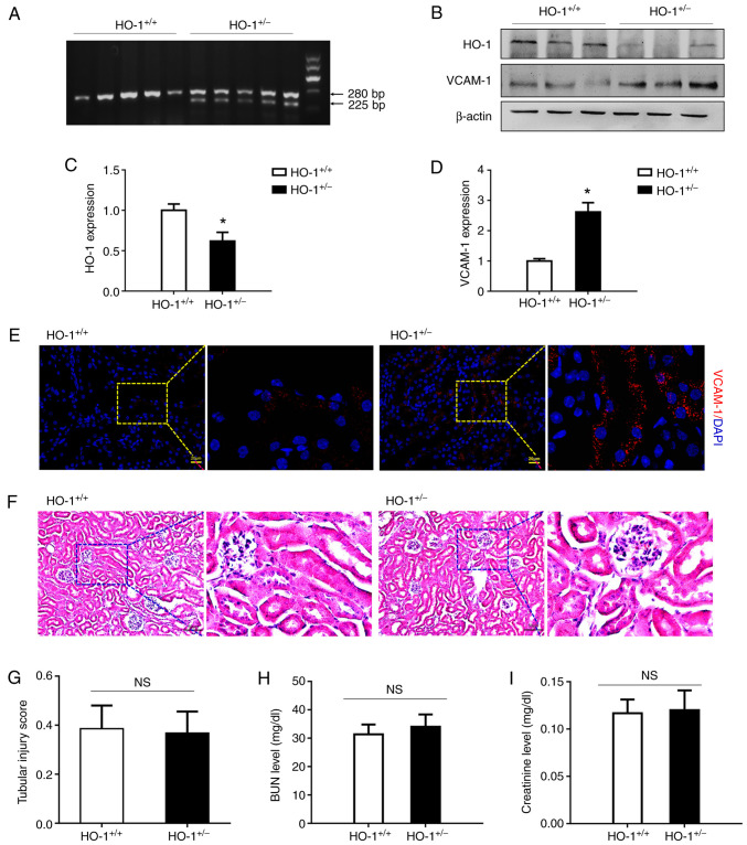
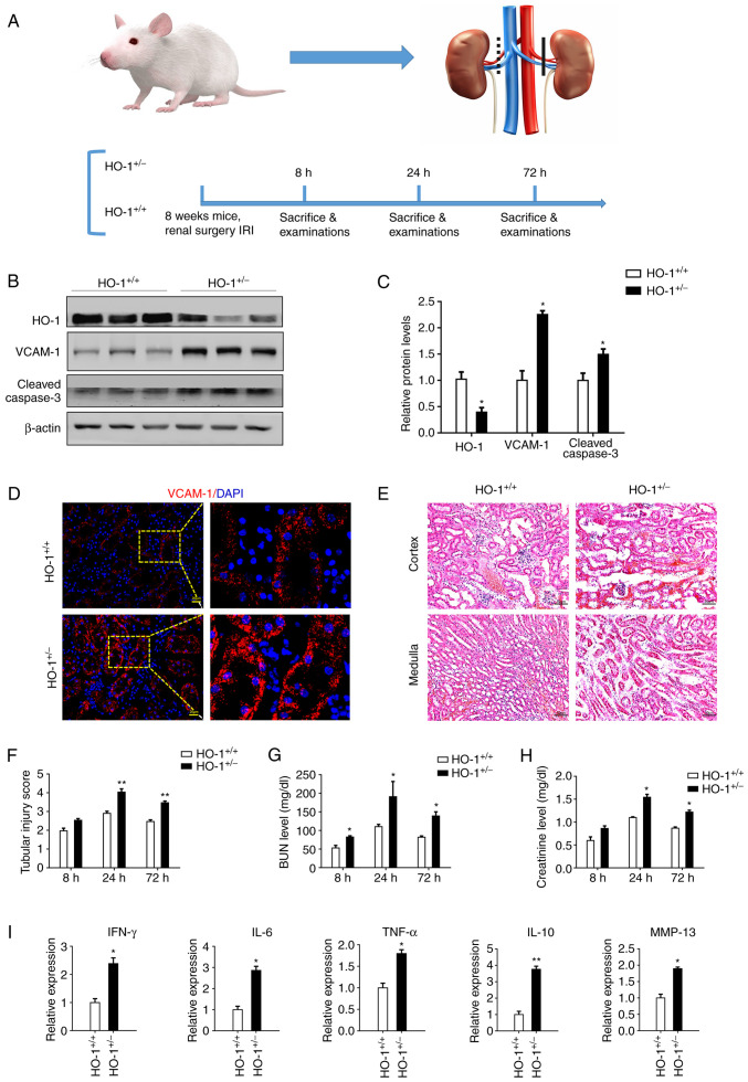
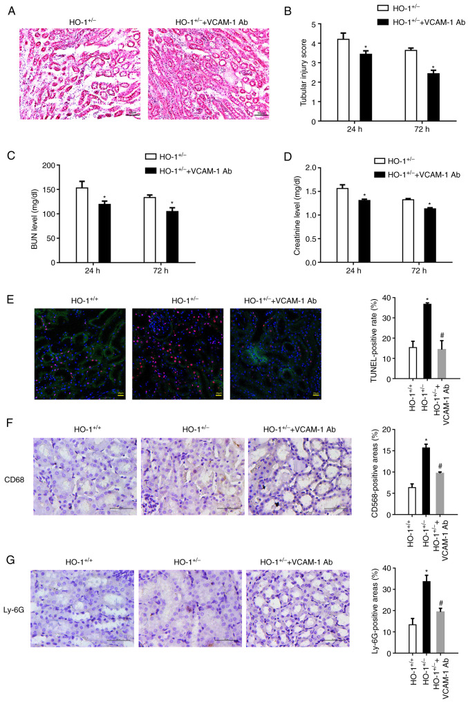
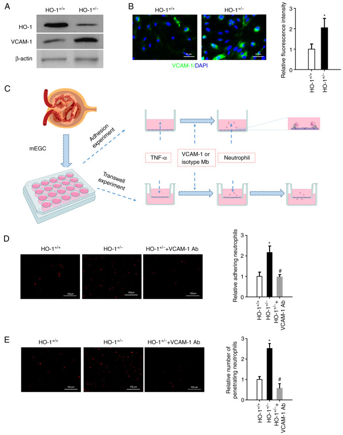
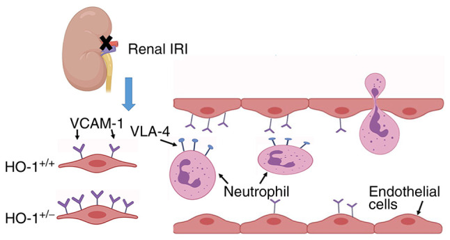
Heme oxygenase‑1 (HO‑1) has been reported to be upregulated following renal ischemia‑reperfusion injury (IRI) and plays a key cytoprotective role; however, the underlying molecular mechanisms of its protective effects remain poorly understood. In the present study, in order to further elucidate the molecular mechanisms underlying the cytoprotective role of HO‑1 in renal IRI, HO‑1+/+ and HO‑1+/‑ mice were subjected to renal ischemia and subsequent reperfusion followed by the analysis of blood urea nitrogen (BUN) and serum creatinine (SCr) levels, the severity of histological changes, HO‑1 and vascular cell adhesion molecule‑1 (VCAM‑1) protein expression, the mRNA expression of inflammatory factors and the effects of VCAM‑1 blockade. The results of the present study demonstrated that the upregulated expression levels of VCAM‑1 in HO‑1+/‑ mice during IRI increased the extent of renal tissue damage and activated the inflammatory response. These effects were subsequently reversed following infusion with an anti‑VCAM‑1 antibody. In addition, the upregulated expression of VCAM‑1 in mouse glomerulus vascular endothelial cells isolated from HO‑1+/‑ mice increased the adhesion and migration of neutrophils, effects which were also reversed upon incubation with an anti‑VCAM‑1 antibody. These results indicated that HO‑1 knockdown may upregulate the expression of VCAM‑1 during renal IRI, resulting in increased neutrophil recruitment and the activation of the inflammatory response, thereby exacerbating renal IRI. The present study thus highlights the regulatory mechanisms of HO‑1 in renal IRI and provides a potential target for the clinical treatment of IRI following renal transplantation.

Keywords: adhesion; heme oxygenase‑1; migration; neutrophil recruitment; renal ischemia‑reperfusion injury; vascular cell adhesion molecule‑1.


Conflict of interest statement

The authors declare that they have no competing interests.

Figures

Figure 1
Figure 1
HO-1+/− knockdown mice exhibit elevated expression levels of VCAM-1 without changes in renal structure and function. (A) Genotype identification of HO+/+ and HO+/− mice by agarose gel electrophoresis. (B) Western blot analysis of the expression levels of HO-1, VCAM-1 and β-actin proteins in the HO-1+/− vs. those in the wild-type mice. (C and D) Densitometric-based quantification of the western blot analysis results shown in panel B for (C) HO-1 and (D) VCAM-1 proteins using ImageJ software. Densitometry values are expressed as the mean ± SD (n=3). *P<0.05 vs. HO-1+/+. (E) Immunohistochemical staining of VCAM-1-expressing cells in the kidneys of the HO-1+/+ and HO-1+/− mice. (F) Representative images of H&E-stained sections of renal tissue in the HO-1+/+ and HO-1+/− mice. (G) Tissue injury was assessed by using the scoring scale from 0 to 5 points (n=3). (H) Serum creatinine concentration in the HO-1+/+ and HO-1+/− mice (n=3). (I) Serum BUN concentration in the HO-1+/+ and HO-1+/− mice (n=3). HO-1, heme oxygenase-1; VCAM-1, vascular cell adhesion molecule-1; BUN, blood urea nitrogen.
Figure 2
Figure 2
Exacerbation of renal IRI in the HO-1+/− mice. (A) Renal ischemia reperfusion model were established by clamping the right renal artery for 60 min and blocking left kidney function in the HO-1+/−, as well as the wild-type mice and tissue harvesting at 8, 24 and 72 h following surgery. (B) Western blot analysis of the expression levels of HO-1, VCAM-1, cleaved caspase-3 and β-actin proteins at 24 h post-IRI in the HO-1+/− vs. those in the wild-type mice. (C) Densitometric-based quantification of the western blot analysis results shown in panel B using ImageJ software. Densitometry values are expressed as the mean ± SD (n=3). *P<0.05 vs. HO-1+/+. (D) Immunohistochemical staining of VCAM-1-expressing cells at 24 h post-IRI in the HO-1+/+ and HO-1+/− mice. (E) Representative images of H&E-stained sections of the cortical and medullary renal tissue showing the structure of the renal tissue. (F) Tissue injury was assessed by using the scoring scale from 0 to 5 points (n=3, **P<0.01 vs. HO-1+/+). (G) Serum BUN concentration at 8, 24 and 72 h post-IRI in the HO-1+/+ and HO-1+/− mice (n=3, *P<0.05 vs. HO-1+/+). (H) Serum creatinine concentration at 8, 24 and 72 post-IRI in the HO-1+/+ and HO-1+/− mice (n=3, *P<0.05 vs. HO-1+/+). (I) Expression levels of IFN-γ, IL-6, TNF-α, IL-10 and MMP-13 in the HO-1+/+ vs. the HO-1+/− group assessed by RT-qPCR (n=3, *P<0.05, **P<0.01 vs. HO-1+/+). IRI, ischemia-reperfusion injury; HO-1, heme oxygenase-1; VCAM-1, vascular cell adhesion molecule-1; BUN, blood urea nitrogen.
Figure 3
Figure 3
VCAM-1 blocking alleviates renal IRI. VCAM-1 antibody was infused into the HO-1+/− knockdown mice through the tail vein to block VCAM-1 expression on the vascular endothelium. (A) Representative images of H&E-stained sections of renal tissue showing its morphology. (B) Extent of the kidney tissue injury was assessed using the 0 to 5-point scoring system (n=3, *P<0.05 vs. HO-1+/−). (C) Serum BUN concentration at 24 and 72 h post-IRI in the HO-1+/+ and HO-1+/− mice (n=3, *P<0.05 vs. HO-1+/−). (D) Serum creatinine concentration at 24 and 72 h post-IRI in the HO-1+/+ and HO-1+/− mice (n=3, *P<0.05 vs. HO-1+/−). (E) Cell death upon IRI was measured using TUNEL assay. The TUNEL-positive rate was analyzed using ImageJ software in the HO-1+/+, HO-1+/− and HO-1+/− + VCAM-1 Ab groups (*P<0.05 vs. HO-1+/+; #P<0.05 vs. HO-1+/−). (F) Immunohistochemical staining and quantification analysis of CD68-expressing cells in mouse kidneys in the HO-1+/+, HO-1+/− and HO-1+/− + VCAM-1 Ab groups (*P<0.05 vs. HO-1+/+; #P<0.05 vs. HO-1+/−). (G) Immunohistochemical staining and quantification of Ly-6G-expressing cells in mouse kidneys of the HO-1+/+, HO-1+/− and HO-1+/− + VCAM-1 Ab groups (*P<0.05 vs. HO-1+/+; #P<0.05 vs. HO-1+/−). IRI, ischemia-reperfusion injury; HO-1, heme oxygenase-1; VCAM-1, vascular cell adhesion molecule-1; BUN, blood urea nitrogen; Ab, antibody.
Figure 4
Figure 4
In vitro blocking of VCAM-1 suppresses neutrophil adhesion and migration through Transwells. mGECs from the HO-1+/− and wild-type mice were isolated. (A) Western blot analysis of the expression levels of HO-1, VCAM-1 and β-actin proteins in the mGECs extracted from the HO-1+/+ and HO-1+/− mice. (B) Immunofluorescence staining of VCAM-1 in the mGECs extracted from the HO-1+/+ and HO-1+/− mice. Relative fluorescent intensity was quantified using ImageJ software (n=3, *P<0.05 vs. HO-1+/+). (C) mGECs were grown in a 96-well plate or Transwell chamber and stimulated with 100 U/ml TNF-α for 4 h. Neutrophils were isolated and labeled with PKH26 to perform adhesion assay or Transwell migration assay. (D) Neutrophils adhered to mGECs were photographed using a fluorescence microscope, and the fluorescence area was quantified using ImageJ software (n=3, *P<0.05 vs. HO-1+/+; #P<0.05 vs. HO-1+/−). (E) Neutrophil migration through mGECs was photographed using a fluorescence microscope, and the fluorescence area was quantified using ImageJ software (n=3, *P<0.05 vs. HO-1+/+; #P<0.05 vs. HO-1+/−). mGECs, mouse glomerular endothelial cells; HO-1, heme oxygenase-1; VCAM-1, vascular cell adhesion molecule-1; Ab, antibody.
Figure 5
Figure 5
Schematic representation of the proposed mechanisms. Neutrophils bind to mGECs through the interaction of VLA-4 and VCAM-1 to activate mGECs. The expression level of VCAM-1 in HO-1+/− knockdown mice is elevated, resulting in excessive neutrophil adhesion and migration through mEGCs, and eventually exacerbating renal IRI. mGECs, mouse glomerular endothelial cells; HO-1, heme oxygenase-1; VLA-4, very late antigen 4; VCAM-1, vascular cell adhesion molecule-1; IRI, ischemia-reperfusion injury.

References

    1. Dong Y, Zhang Q, Wen J, Chen T, He L, Wang Y, Yin J, Wu R, Xue R, Li S, et al. Ischemic duration and frequency determines AKI-to-CKD progression monitored by dynamic changes of tubular biomarkers in IRI mice. Front Physiol. 2019;10:153. doi: 10.3389/fphys.2019.00153. - DOI - PMC - PubMed
    1. Sheashaa H, Lotfy A, Elhusseini F, Aziz AA, Baiomy A, Awad S, Alsayed A, El-Gilany AH, Saad MA, Mahmoud K, et al. Protective effect of adipose-derived mesenchymal stem cells against acute kidney injury induced by ischemia-reperfusion in Sprague-Dawley rats. Exp Ther Med. 2016;11:1573–1580. doi: 10.3892/etm.2016.3109. - DOI - PMC - PubMed
    1. Cheng Q, Wang L. LncRNA XIST serves as a ceRNA to regulate the expression of ASF1A, BRWD1M, and PFKFB2 in kidney transplant acute kidney injury via sponging hsa-miR-212-3p and hsa-miR-122-5p. Cell Cycle. 2020;19:290–299. doi: 10.1080/15384101.2019.1707454. - DOI - PMC - PubMed
    1. Yang C, Qi R, Yang B. Pathogenesis of chronic allograft dysfunction progress to renal fibrosis. Adv Exp Med Biol. 2019;1165:101–116. doi: 10.1007/978-981-13-8871-2_6. - DOI - PubMed
    1. Zhang A, Wan B, Jiang D, Wu Y, Ji P, Du Y, Zhang G. The cytoprotective enzyme heme oxygenase-1 suppresses pseudorabies virus replication in vitro. Front Microbiol. 2020;11:412. doi: 10.3389/fmicb.2020.00412. - DOI - PMC - PubMed

MeSH terms