. 2021 Oct 12;203(21):e0019621.
doi: 10.1128/JB.00196-21. Epub 2021 Aug 9.

The Corynebacterium diphtheriae HbpA Hemoglobin-Binding Protein Contains a Domain That Is Critical for Hemoprotein Binding, Cellular Localization, and Function

Affiliations

The Corynebacterium diphtheriae HbpA Hemoglobin-Binding Protein Contains a Domain That Is Critical for Hemoprotein Binding, Cellular Localization, and Function

Lindsey R Lyman et al. J Bacteriol. .

Abstract

The acquisition of hemin iron from hemoglobin-haptoglobin (Hb-Hp) by Corynebacterium diphtheriae requires the iron-regulated surface proteins HtaA, ChtA, and ChtC and the recently identified Hb-Hp-binding protein, HbpA. We previously showed that a purified form of HbpA (HbpA-S), lacking the C-terminal region, was able to bind Hb-Hp. In this study, we show that the C-terminal region of HbpA significantly enhances binding to Hb-Hp. A purified form of HbpA that includes the C-terminal domain (HbpA-FL) exhibits much stronger binding to Hb-Hp than HbpA-S. Size exclusion chromatography (SEC) showed that HbpA-FL as well as HtaA-FL, ChtA-FL, and ChtC-FL exist as high-molecular-weight complexes, while HbpA-S is present as a monomer, indicating that the C-terminal region is required for formation of large aggregates. Growth studies showed that expression of HbpA-FL in the ΔhbpA mutant restored wild-type levels of growth in low-iron medium that contained Hb-Hp as the sole iron source, while HbpA-S failed to complement the ΔhbpA mutant. Protein localization studies in C. diphtheriae showed that HbpA-FL is present in both the supernatant and membrane fractions and that the C-terminal region is required for membrane anchoring. Purified HbpA-FL was able to enhance growth of the ΔhbpA mutant when added to culture medium that contained Hb-Hp as a sole iron source, suggesting that secreted HbpA is involved in the use of hemin iron from Hb-Hp. These studies extend our understanding of this novel Hb-Hp binding protein in this important human pathogen. IMPORTANCE Hemoproteins, such as Hb, are an abundant source of iron in humans and are proposed to be required by numerous pathogens to cause disease. In this report, we expand on our previous studies in further defining the role of HbpA in hemin iron acquisition in C. diphtheriae. HbpA is unique to C. diphtheriae and appears to function unlike any previously described bacterial iron-regulated Hb- or Hb-Hp-binding protein. HbpA is both secreted and present in the membrane and exists as a large aggregate that enhances its ability to bind Hb-Hp and promote hemin iron uptake. Current studies with HbpA will increase our understanding of iron transport systems in C. diphtheriae.

Keywords: Corynebacterium; diphtheria; hemin; hemoglobin; iron transport.


Figures

FIG 1
FIG 1
(A) Genetic map of wild-type (WT) hbpA and expression constructs HbpA-FL and HbpA-S. The expression constructs lack the N-terminal signal sequence (SS), and HbpA-S also lacks the C-terminal sequence (underlined sequence below map). The C-terminal sequence includes the transmembrane domain (TM; sequence indicated in red) and positively charged residues (+; bold text). (B) Purified recombinant strep-tag II-tagged HbpA-S and HbpA-FL were separated by SDS-PAGE and then either stained with Coomassie blue (upper panel) or incubated in situ with Hb-Hp, which was then detected by anti-Hp antibodies (lower panel). The protein amount loaded for each lane is indicated below the gel. A representative experiment is shown.
FIG 2
FIG 2
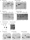
Hb-Hp binding to C. diphtheriae hemin transport proteins. (A) The FL and S derivatives of various recombinant proteins were assessed for binding to Hb-Hp. Proteins were separated by SDS-PAGE (4 to 15% gradient gel) and were stained with Coomassie blue (left panel) or examined for binding to Hb-Hp using an in situ method (right panel). The amount of protein loaded for each lane is indicated below the gel. ChtA Term was used as a negative binding control. Note reduced quantities (micrograms loaded) of HbpA-FL. (B) The same proteins used in panel A were separated using 4 to 15% gradient native gels. The presence (+) or absence (−) of 0.1% Tween 20 is indicated for FL proteins. (C) Comparison of supernatant (Sup) from C. diphtheriae 1737Δ3 grown in low-iron (0.25 μM FeCl3) mPGT medium and purified recombinant HbpA-FL separated on a native 4 to 15% gel. Left panel, anti-HbpA Western blot; right panel, Hb-Hp in situ binding detected with anti-Hp antibodies. Plus and minus indicate the presence and absence, respectively, of 0.1% Tween 20. (D) Comparison of Hb and Hb-Hp binding between the indicated proteins. Proteins were separated by SDS-PAGE (4 to 15% gradient gel). The amount of protein loaded in each lane is indicated below the gel. Binding to Hb and Hb-Hp was done in situ as described in Materials and Methods. Representative experiments are shown.
FIG 3
FIG 3
SEC analysis of C. diphtheriae hemin transport protein HbpA. (A) SEC traces showing purified recombinant HbpA-S, HbpA-FL, and HbpA-FL preincubated with increasing amounts of Tween 20 (Tw). Higher retention times indicate smaller-molecular-weight species. Retention times of molecular weight standards (Bio-Rad gel filtration standards) are indicated by dotted lines. (B) SEC molecular weight standards and purified HbpA-FL protein were analyzed using a Superose 6 increase 3.2/300 chromatography system. The elution trace shows the column retention times of HbpA-FL and three high-molecular-weight standards: blue dextran (BD), thyroglobulin (Thy), and ferritin (Fer). All graphs show a representative sample from at least three replicates.
FIG 4
FIG 4
HbpA-FL, HbpA-S, and IsdH-N2N3 were assessed for their ability to bind Hb (A) and Hb-Hp (B) by ELISA. Purified Hb or Hb-Hp was bound to a microtiter plate, washed, and then incubated with the indicated strep-tag II-tagged proteins. Binding was detected by anti-strep tag II antibody, and absorbance was measured at 405 nm. HbpA-FL was tested at significantly lower concentrations than the other proteins due to high binding affinity (see insets for HbpA-FL). Graphs show the means ± standard deviations of the results of three experiments.
FIG 5
FIG 5
Purified recombinant proteins at 5 μM were incubated with 5 μM heme for 20 min at room temperature and then scanned for absorbance from 200 to 700 nm. The scan was baselined against heme in the absence of protein. HtaA-S shows a clear Soret peak at 407 nm and a small peak between 625 and 630 nm (see inset). HbpA-FL shows a peak at 412 nm; HbpA-S and ChtA Term (a non-binding region of the ChtA protein) show minimal to no peak in the 400-nm region. The graph shows the average results of three replicates. GraphPad Prism 9.0.0 was used to apply second-order smoothing (using 6 neighbors) to the data to enhance visualization.
FIG 6
FIG 6
Growth assays of C. diphtheriae 1737 WT and various mutants to assess the impact of the C-terminal region on growth with Hb-Hp. Bacteria were grown in mPGT medium with Hb-Hp as the sole iron source, and overnight growth was measured by OD600. Plasmid pKN (vector pKN2.6Z) and various full-length (FL) or short (S) complementation clones constructed using pKN2.6Z are indicated below the graph. Error bars show means with standard deviations of the results of three experiments. ****, P <0.0001; ***, P <0.001; *, P <0.05; ns, no significant difference.
FIG 7
FIG 7
Cell fractionation studies with C. diphtheriae 1737 WT and 1737ΔhbpA carrying the cloned genes for hbpA-FL or hbpA-S. Bacteria were grown in low-iron (0.25 μM FeCl3) mPGT medium and harvested after overnight growth. Protein fractions include the supernatant (S), total cell lysate (T), cytosol (C), and membrane (M). The left panel shows a Western blot probed with anti-HbpA, and the right panel shows a Coomassie blue-stained gel. pKN indicates vector pKN2.6Z; all clones were constructed using pKN2.6Z. A representative experiment is shown.
FIG 8
FIG 8
A whole-cell ELISA was used to detect the presence of HbpA, HtaA, and ChtA on the surface of C. diphtheriae strains that were grown in low-iron (0.25 μM FeCl3) mPGT medium. Following overnight growth, cell density was normalized by OD600 and bacteria were bound to 96-well high-protein-binding plates, and then expression was assessed using antibodies to the proteins of interest. Strains tested include C. diphtheriae 1737 WT, 1737ΔhbpAhbpA), or 1737Δ4 (Δ4), which is deleted for hbpA, htaA, chtA, and chtC. pKN indicates vector pKN2.6Z; all clones were constructed using pKN2.6Z. The full-length (FL) or short (S) hbpA cloned genes are indicated below the graph. DtxR is a cytosolic protein that was used as a negative control. The graph shows the means ± standard deviations of the results of three experiments.
FIG 9
FIG 9
Detergent treatment was used to extract cell-associated proteins that are loosely anchored to the bacterial surface. C. diphtheriae 1737 wild-type cells were grown in low-iron (0.25 μM) mPGT medium and harvested after overnight growth. Culture supernatant fractions (S) were collected following growth. Cell pellets were either treated immediately with muramidase and Sarkosyl to obtain the total cell lysate (T) containing all cellular proteins or first incubated in RIPA buffer to isolate weakly associated proteins (R) and then treated with muramidase and Sarkosyl to isolate the remaining cellular proteins (C). Fractions were separated by SDS-PAGE, and Western blots were probed using antibodies specific to HbpA, ChtC, IutA, and DtxR as indicated. IutA, a membrane-bound lipoprotein, and DtxR, a cytosolic protein, served as controls. Fractions run on SDS-PAGE were also stained using Coomassie blue (right panel).
FIG 10
FIG 10
The addition of HbpA-FL protein to culture medium enhances growth of the hbpA mutant. (A) C. diphtheriae 1737ΔhbpA cells were grown in mPGT medium with Hb-Hp as the sole iron source, and 5 μg of purified recombinant HbpA-S (rHbpA-S) or rHbpA-FL was added to the growth medium as indicated. Bacterial density was assessed by measuring the OD600 of terminal growth cultures. Controls that contained no added protein included WT/pKN (vector) and 1737ΔhbpA with plasmid pKN-hbpA-FL or pKN. Growth data show means with standard deviations of the results of at least three replicates. ****, P <0.0001; **, P <0.01; *, P <0.05; ns, no significant difference. (B) Western blot analysis of cultures used in panel A. Culture densities were normalized by OD and harvested by centrifugation, and the supernatant (S) was collected. The remaining bacteria were then treated with lysozyme and Sarkosyl to obtain the total cell fraction (T), and proteins were separated by SDS-PAGE. Protein was detected using anti-HbpA antibodies from the samples indicated above the gel. The Western blot is a representative experiment.

References

    1. Zasada AA. 2015. Corynebacterium diphtheriae infections currently and in the past. Przegl Epidemiol 69:439–444, 569-574. - PubMed
    1. Hadfield TL, McEvoy P, Polotsky Y, Tzinserling VA, Yakovlev AA. 2000. The pathology of diphtheria. J Infect Dis 181(Suppl 1):S116–S120. 10.1086/315551. - DOI - PubMed
    1. Collier RJ. 2001. Understanding the mode of action of diphtheria toxin: a perspective on progress during the 20th century. Toxicon 39:1793–1803. 10.1016/s0041-0101(01)00165-9. - DOI - PubMed
    1. Holmes RK. 2000. Biology and molecular epidemiology of diphtheria toxin and the tox gene. J Infect Dis 181(Suppl 1):S156–S167. 10.1086/315554. - DOI - PubMed
    1. Boyd J, Oza MN, Murphy JR. 1990. Molecular cloning and DNA sequence analysis of a diphtheria tox iron-dependent regulatory element (dtxR) from Corynebacterium diphtheriae. Proc Natl Acad Sci USA 87:5968–5972. 10.1073/pnas.87.15.5968. - DOI - PMC - PubMed

Publication types

MeSH terms