. 2021 Aug 9;17(8):e1009802.
doi: 10.1371/journal.ppat.1009802. eCollection 2021 Aug.

Modern Acinetobacter baumannii clinical isolates replicate inside spacious vacuoles and egress from macrophages

Affiliations

Modern Acinetobacter baumannii clinical isolates replicate inside spacious vacuoles and egress from macrophages

Gabriela Sycz et al. PLoS Pathog. .

Abstract

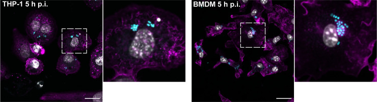
Multidrug-resistant Acinetobacter baumannii infections are increasing at alarming rates. Therefore, novel antibiotic-sparing treatments to combat these A. baumannii infections are urgently needed. The development of these interventions would benefit from a better understanding of this bacterium's pathobiology, which remains poorly understood. A. baumannii is regarded as an extracellular opportunistic pathogen. However, research on Acinetobacter has largely focused on common lab strains, such as ATCC 19606, that have been isolated several decades ago. These strains exhibit reduced virulence when compared to recently isolated clinical strains. In this work, we demonstrate that, unlike ATCC 19606, several modern A. baumannii clinical isolates, including the recent clinical urinary isolate UPAB1, persist and replicate inside macrophages within spacious vacuoles. We show that intracellular replication of UPAB1 is dependent on a functional type I secretion system (T1SS) and pAB5, a large conjugative plasmid that controls the expression of several chromosomally-encoded genes. Finally, we show that UPAB1 escapes from the infected macrophages by a lytic process. To our knowledge, this is the first report of intracellular growth and replication of A. baumannii. We suggest that intracellular replication within macrophages may contribute to evasion of the immune response, dissemination, and antibiotic tolerance of A. baumannii.


Conflict of interest statement

The authors have declared that no competing interests exist.

Figures

Fig 1
Fig 1. UPAB1 resides in spacious vacuoles within macrophages.
Transmission electron microscopic images of J774A.1 macrophages infected with UPAB1 (A) at 5 (left panel) and 7 h p.i. (right panel) Bars: 2 μm. (B) The UPAB1 containing vacuole can have a single (left panel, white arrow) or double membrane (right panel, white arrow). Bars: 500 nm.
Fig 2
Fig 2. UPAB1 but not 19606 replicates in macrophages.
(A) J774A.1 macrophages were infected with the indicated strains, and after phagocytosis, extracellular bacteria were eliminated with antibiotics. After different time points, total numbers of intracellular CFU were determined. *p < 0.05. (B) Representative images of infected cells at the indicated time points. Samples were stained for cell nuclei (grey), actin (magenta) and Acinetobacter GFP (cyan). Insets show a higher magnification of the area indicated by the white box. Individual channels are presented in S3 Fig. Bars: 20 μm. (C) Quantification of bacteria per vacuole at different times p.i. Each symbol represents an individual vacuole, horizontal line represents the median. At least 200 infected cells were analyzed for each time point for three independent experiments.
Fig 3
Fig 3. UPAB1 replicates in multiple host cell lines.
Representative images of infected THP-1 (left panels) or BMDM (right panels) cells at 5 h p.i. Samples were stained for cell nuclei (grey), actin (magenta) and UPAB1 GFP (cyan). Insets show a higher magnification of the area indicated by the white box. Individual channels are presented in S4 Fig. Bars: 20 μm.
Fig 4
Fig 4. Recent clinical isolates replicate in macrophages.
(A) J774A.1 macrophages were infected with the indicated strains and, after phagocytosis, extracellular bacteria were eliminated with antibiotics. After different time points, total numbers of intracellular CFU were determined. Data represent mean and standard deviation values for 3 independent experiments. (B) Representative images of infected cells with the indicated strains. Samples were stained for cell nuclei (grey), actin (magenta) and Acinetobacter GFP (cyan). Insets show a higher magnification of the area indicated by the white box. Individual channels are presented in S5 Fig. Bars: 20 μm.
Fig 5
Fig 5. UPAB1 escapes the macrophages by a lytic process.
J774A.1 macrophages were infected with UPAB1. (A) At different time points, antibiotic containing medium was replaced by antibiotic-free medium. CFU in supernatants were determined and the ratio of released to intracellular bacteria was calculated. Data represent mean and standard deviation values for 3 independent experiments. (B) Images from time-lapse microscopy showing UPAB1 egress from the invaded macrophages. J774A.1 cells were infected with UPAB1 GFP (green fluorescence). At 7 h p.i. images were taken every 10 min and analyzed with ImageJ software. The white arrow indicates the infected cell. Bars: 20 μm. (C) LDH activity in the supernatant of infected macrophages was measured at 24 h p.i. Percentage of cytotoxicity was calculated as the activity of released LDH relative to total LDH activity. The mean ± S.D. for two independent experiments is shown. **** p < 0.0001.
Fig 6
Fig 6. pAB5 and T1SS are required for UPAB1 intracellular replication.
(A) J774A.1 macrophages were infected with the indicated strains. UPAB1p-, UPAB1pAB3+ and UPAB1pAB5Δh-ns strains were not able to replicate. Data represents mean and standard deviation values for 3 independent experiments. *p < 0.05. (B) Genetic organization of the T1SSa and T1SSb loci. (C) Individual and double T1SS mutants have a defect in intracellular replication in J774A.1 macrophages. Data represents mean and standard deviation values, for 3 independent experiments. *p < 0.05.

References

    1. Weiner LM, Webb AK, Limbago B, Dudeck MA, Patel J, Kallen AJ, et al.. Antimicrobial-Resistant Pathogens Associated With Healthcare-Associated Infections: Summary of Data Reported to the National Healthcare Safety Network at the Centers for Disease Control and Prevention, 2011–2014. Infect Control Hosp Epidemiol. 2016;37: 1288–1301. doi: 10.1017/ice.2016.174 - DOI - PMC - PubMed
    1. Valencia R, Arroyo LA, Conde M, Aldana JM, Torres M-J, Fernández-Cuenca F, et al.. Nosocomial outbreak of infection with pan-drug-resistant Acinetobacter baumannii in a tertiary care university hospital. Infect Control Hosp Epidemiol. 2009;30: 257–63. doi: 10.1086/595977 - DOI - PubMed
    1. Tacconelli E, Magrini N, Kahlmeter G, Singh N. Global Priority List of Antibiotic-Resistant Bacteria To Guide Research, Discovery, and Development of New Antibiotics. 2017.
    1. Centers for Disease Control U. Antibiotic Resistance Threats in the United States, 2019. [cited 8 Apr 2021]. doi: 10.15620/cdc:82532 - DOI
    1. Di Venanzio G, Flores-Mireles AL, Calix JJ, Haurat MF, Scott NE, Palmer LD, et al.. Urinary tract colonization is enhanced by a plasmid that regulates uropathogenic Acinetobacter baumannii chromosomal genes. Nat Commun. 2019;10: 1100. doi: 10.1038/s41467-019-08917-4 - DOI - PMC - PubMed

Publication types

MeSH terms

Substances

LinkOut - more resources