

Acute disseminated encephalomyelitis and COVID-19: A systematic synthesis of worldwide cases
- PMID: 34371208
- PMCID: PMC8313793
- DOI: 10.1016/j.jneuroim.2021.577674
Acute disseminated encephalomyelitis and COVID-19: A systematic synthesis of worldwide cases
Abstract
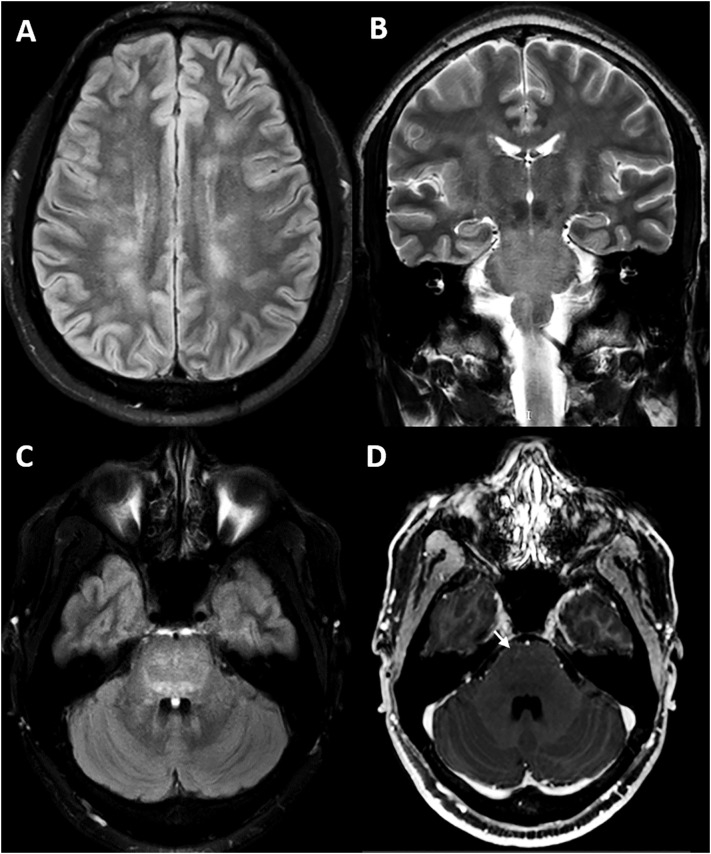
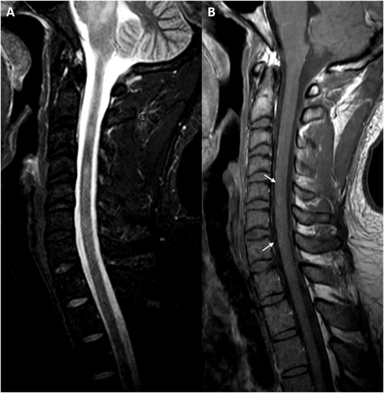
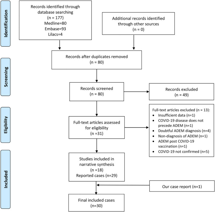
Acute disseminated encephalomyelitis (ADEM) has been reported after coronavirus disease 2019 (COVID-19). In this review, we systematically included worldwide reported cases on this association. We included 30 case reports (pediatric and adults) and explored epidemiological and clinical evidence. We described time to diagnosis, clinical, imaging, and laboratory features, response to treatment regimens, and differences regarding severity. Also, an original case report was presented. Neurologists must be alert to the occurrence of multifocal neurological symptoms with or without encephalopathy in patients recovered from COVID-19. Timely MRI studies should be performed to establish the diagnosis and to consider early corticosteroid-based treatment.
Keywords: Acute disseminated encephalomyelitis (ADEM); Autoimmune disease; Central nervous system (CNS); Coronavirus disease 2019 (COVID-19); Demyelinating disease; SARS-Cov-2.
Copyright © 2021 Elsevier B.V. All rights reserved.
Figures
Similar articles
-
Complex movement disorders in SARS-CoV-2 infection induced acute disseminated encephalomyelitis.J Neuroimmunol. 2021 Sep 15;358:577655. doi: 10.1016/j.jneuroim.2021.577655. Epub 2021 Jun 30. J Neuroimmunol. 2021. PMID: 34265625 Free PMC article.
-
SUCCESSFUL TREATMENT OF ACUTE DISSEMINATED ENCEPHALOMYELITIS (ADEM) BY PROMPT USAGE OF IMMUNOGLOBULINS - CASE REPORT AND REVIEW OF THE LITERATURE.Acta Clin Croat. 2019 Mar;58(1):173-179. doi: 10.20471/acc.2019.58.01.22. Acta Clin Croat. 2019. PMID: 31363340 Free PMC article.
-
Acute Disseminated Encephalomyelitis and Acute Encephalitis Following Vaccination Against SARS-CoV-2: Two Case Reports and Review of Literature.Fortschr Neurol Psychiatr. 2023 Jul;91(7-08):319-325. doi: 10.1055/a-2029-4405. Epub 2023 Mar 17. Fortschr Neurol Psychiatr. 2023. PMID: 36931294 Review. English.
-
Acute haemorrhagic necrotizing encephalopathy and inflammatory demyelinating encephalopathy associated with COVID-19 in adults in Southern China.BMC Infect Dis. 2025 Aug 7;25(1):990. doi: 10.1186/s12879-025-11404-5. BMC Infect Dis. 2025. PMID: 40775306 Free PMC article.
-
Acute Disseminated Encephalomyelitis and Acute Hemorrhagic Leukoencephalitis Following COVID-19: Systematic Review and Meta-synthesis.Neurol Neuroimmunol Neuroinflamm. 2021 Aug 27;8(6):e1080. doi: 10.1212/NXI.0000000000001080. Print 2021 Nov. Neurol Neuroimmunol Neuroinflamm. 2021. PMID: 34452974 Free PMC article.
Cited by
-
The Occurrence of Acute Disseminated Encephalomyelitis in SARS-CoV-2 Infection/Vaccination: Our Experience and a Systematic Review of the Literature.Vaccines (Basel). 2023 Jul 10;11(7):1225. doi: 10.3390/vaccines11071225. Vaccines (Basel). 2023. PMID: 37515041 Free PMC article. Review.
-
Acute Disseminated Encephalomyelitis Following COVID-19 Infection.Cureus. 2023 Jan 4;15(1):e33365. doi: 10.7759/cureus.33365. eCollection 2023 Jan. Cureus. 2023. PMID: 36751218 Free PMC article.
-
Encephalopathy as a prognostic factor in adults with acute disseminated encephalomyelitis following COVID-19.J Neurol. 2022 May;269(5):2293-2300. doi: 10.1007/s00415-021-10947-2. Epub 2022 Jan 3. J Neurol. 2022. PMID: 34978621 Free PMC article.
-
COVID-19 and neurologic manifestations: a synthesis from the child neurologist's corner.World J Pediatr. 2022 Jun;18(6):373-382. doi: 10.1007/s12519-022-00550-4. Epub 2022 Apr 27. World J Pediatr. 2022. PMID: 35476245 Free PMC article. Review.
-
Acute disseminated encephalomyelitis with bilateral optic neuritis following ChAdOx1 COVID-19 vaccination.BMC Neurol. 2022 Feb 12;22(1):54. doi: 10.1186/s12883-022-02575-8. BMC Neurol. 2022. PMID: 35151258 Free PMC article.
References
Publication types
MeSH terms
Substances
LinkOut - more resources
Full Text Sources
Medical
Miscellaneous
